5VAS | pdb_00005vas

Pekin duck egg lysozyme isoform III (DEL-III), orthorhombic form


Experimental Data Snapshot

  • Method: X-RAY DIFFRACTION
  • Resolution: 1.40 Å
  • R-Value Free: 
    0.169 (Depositor), 0.180 (DCC) 
  • R-Value Work: 
    0.144 (Depositor), 0.151 (DCC) 
  • R-Value Observed: 
    0.145 (Depositor) 

Starting Model: experimental
View more details

wwPDB Validation 3D Report Full Report

Validation slider image for 5VAS

This is version 1.4 of the entry. See complete history

Literature

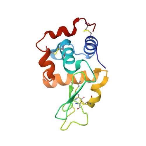
Structural basis of antigen recognition: crystal structure of duck egg lysozyme.

Langley, D.B.Crossett, B.Schofield, P.Jackson, J.Zeraati, M.Maltby, D.Christie, M.Burnett, D.Brink, R.Goodnow, C.Christ, D.

(2017) Acta Crystallogr D Struct Biol 73: 910-920

  • DOI: https://doi.org/10.1107/S2059798317013730
  • Primary Citation Related Structures: 
    5V8G, 5V92, 5V94, 5VAS

  • PubMed Abstract: 

    Duck egg lysozyme (DEL) is a widely used model antigen owing to its capacity to bind with differential affinity to anti-chicken egg lysozyme antibodies. However, no structures of DEL have so far been reported, and the situation had been complicated by the presence of multiple isoforms and conflicting reports of primary sequence. Here, the structures of two DEL isoforms from the eggs of the commonly used Pekin duck (Anas platyrhynchos) are reported. Using structural analyses in combination with mass spectrometry, non-ambiguous DEL primary sequences are reported. Furthermore, the structures and sequences determined here enable rationalization of the binding affinity of DEL for well documented landmark anti-lysozyme antibodies.


  • Organizational Affiliation
    • Immunology Division, Garvan Institute of Medical Research, 384 Victoria Street, Darlinghurst, NSW 2010, Australia.

Macromolecule Content 

  • Total Structure Weight: 30 kDa 
  • Atom Count: 2,404 
  • Modeled Residue Count: 258 
  • Deposited Residue Count: 258 
  • Unique protein chains: 1

Macromolecules

Find similar proteins by:|  3D Structure
Entity ID: 1
MoleculeChains  Sequence LengthOrganismDetailsImage
Lysozyme
A, B
129Anas platyrhynchosMutation(s): 0 
EC: 3.2.1.17

Small Molecules

Ligands 2 Unique
IDChains Name / Formula / InChI Key2D Diagram3D Interactions
PO4

Query on PO4


Download:Ideal Coordinates CCD File
C [auth A],
D [auth A],
H [auth B]
PHOSPHATE ION
O4 P
NBIIXXVUZAFLBC-UHFFFAOYSA-K
GOL

Query on GOL


Download:Ideal Coordinates CCD File
E [auth A],
F [auth A],
G [auth A],
I [auth B],
J [auth B]
GLYCEROL
C3 H8 O3
PEDCQBHIVMGVHV-UHFFFAOYSA-N

Experimental Data & Validation

Experimental Data

  • Method: X-RAY DIFFRACTION
  • Resolution: 1.40 Å
  • R-Value Free:  0.169 (Depositor), 0.180 (DCC) 
  • R-Value Work:  0.144 (Depositor), 0.151 (DCC) 
  • R-Value Observed: 0.145 (Depositor) 
Space Group: P 21 21 21
Unit Cell:
Length ( Å )Angle ( ˚ )
a = 44.005α = 90
b = 58.35β = 90
c = 107.48γ = 90
Software Package:
Software NamePurpose
MOSFLMdata collection
Aimlessdata scaling
PHASERphasing
PDB_EXTRACTdata extraction
MOSFLMdata reduction
REFMACrefinement

Structure Validation

View Full Validation Report


Entry History 

& Funding Information

Deposition Data


Funding OrganizationLocationGrant Number
National Health and Medical Research Council (NHMRC, Australia)Australia--

Revision History  (Full details and data files)

  • Version 1.0: 2018-04-18
    Type: Initial release
  • Version 1.1: 2019-05-01
    Changes: Data collection, Database references
  • Version 1.2: 2020-01-08
    Changes: Author supporting evidence
  • Version 1.3: 2023-10-04
    Changes: Data collection, Database references, Refinement description
  • Version 1.4: 2024-11-06
    Changes: Structure summary