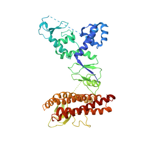
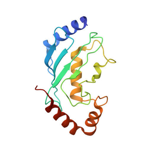
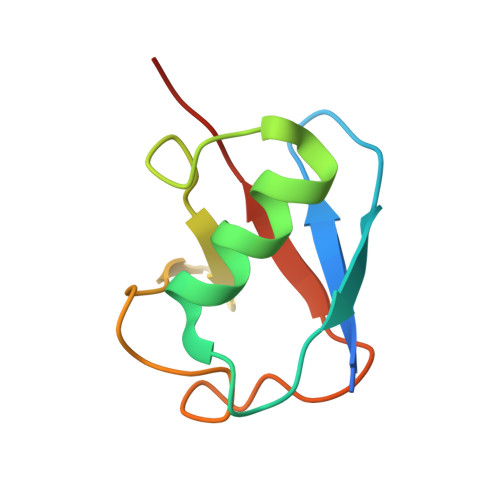
5UDH | pdb_00005udh

HHARI/ARIH1-UBCH7~Ubiquitin


Experimental Data Snapshot

  • Method: X-RAY DIFFRACTION
  • Resolution: 3.24 Å
  • R-Value Free: 
    0.279 (Depositor), 0.277 (DCC) 
  • R-Value Work: 
    0.222 (Depositor), 0.224 (DCC) 
  • R-Value Observed: 
    0.225 (Depositor) 

wwPDB Validation 3D Report Full Report

Validation slider image for 5UDH

This is version 1.2 of the entry. See complete history. 

Literature

Structural Studies of HHARI/UbcH7Ub Reveal Unique E2Ub Conformational Restriction by RBR RING1.

Dove, K.K., Olszewski, J.L., Martino, L., Duda, D.M., Wu, X.S., Miller, D.J., Reiter, K.H., Rittinger, K., Schulman, B.A., Klevit, R.E.

(2017) Structure 25: 890-900.e5

  • DOI: https://doi.org/10.1016/j.str.2017.04.013
  • Primary Citation Related Structures: 
    5UDH

  • PubMed Abstract: 

    RING-between-RING (RBR) E3s contain RING1 domains that are structurally similar yet mechanistically distinct from canonical RING domains. Both types of E3 bind E2∼ubiquitin (E2∼Ub) via their RINGs but canonical RING E3s promote closed E2∼Ub conformations required for direct Ub transfer from the E2 to substrate, while RBR RING1s promote open E2∼Ub to favor Ub transfer to the E3 active site. This different RING/E2∼Ub conformation determines its direct target, which for canonical RING E3s is typically a substrate or substrate-linked Ub, but is the E3 active-site cysteine in the case of RBR-type E3s. Here we show that a short extension of HHARI RING1, namely Zn 2+ -loop II, not present in any RING E3s, acts as a steric wedge to disrupt closed E2∼Ub, providing a structural explanation for the distinctive RING1-dependent conformational restriction mechanism utilized by RBR E3s.


  • Organizational Affiliation: 
    • Department of Biochemistry, University of Washington, 1705 Northeast Pacific Street, Seattle, WA 98195, USA.

Macromolecule Content 

  • Total Structure Weight: 157.62 kDa 
  • Atom Count: 9,178 
  • Modeled Residue Count: 1,172 
  • Deposited Residue Count: 1,334 
  • Unique protein chains: 3

Macromolecules

Find similar proteins by:|  3D Structure
Entity ID: 1
MoleculeChains  Sequence LengthOrganismDetailsImage
E3 ubiquitin-protein ligase ARIH1A,
C [auth B]
470Homo sapiensMutation(s): 0 
Gene Names: ARIH1, ARI, MOP6, UBCH7BP, HUSSY-27
EC: 2.3.2 (PDB Primary Data), 2.3.2.31 (UniProt)
UniProt & NIH Common Fund Data Resources
Find proteins for Q9Y4X5 (Homo sapiens)
Explore Q9Y4X5 
Go to UniProtKB:  Q9Y4X5
PHAROS:  Q9Y4X5
GTEx:  ENSG00000166233 
Entity Groups
Sequence Clusters30% Identity50% Identity70% Identity90% Identity95% Identity100% Identity
UniProt GroupQ9Y4X5
Sequence Annotations
Expand
Reference Sequence
Find similar proteins by:|  3D Structure
Entity ID: 2
MoleculeChains  Sequence LengthOrganismDetailsImage
Ubiquitin-conjugating enzyme E2 L3B [auth C],
D
156Homo sapiensMutation(s): 1 
Gene Names: UBE2L3, UBCE7, UBCH7
EC: 2.3.2.23
UniProt & NIH Common Fund Data Resources
Find proteins for P68036 (Homo sapiens)
Explore P68036 
Go to UniProtKB:  P68036
PHAROS:  P68036
GTEx:  ENSG00000185651 
Entity Groups
Sequence Clusters30% Identity50% Identity70% Identity90% Identity95% Identity100% Identity
UniProt GroupP68036
Sequence Annotations
Expand
Reference Sequence
Find similar proteins by:|  3D Structure
Entity ID: 3
MoleculeChains  Sequence LengthOrganismDetailsImage
Ubiquitin C variant82Homo sapiensMutation(s): 0 
UniProt & NIH Common Fund Data Resources
Find proteins for P0CG48 (Homo sapiens)
Explore P0CG48 
Go to UniProtKB:  P0CG48
PHAROS:  P0CG48
GTEx:  ENSG00000150991 
Entity Groups
Sequence Clusters30% Identity50% Identity70% Identity90% Identity95% Identity100% Identity
UniProt GroupP0CG48
Sequence Annotations
Expand
Reference Sequence

Small Molecules

Ligands 1 Unique
IDChains Name / Formula / InChI Key2D Diagram3D Interactions
ZN

Query on ZN


Download:Ideal Coordinates CCD File
F [auth A]
G [auth A]
H [auth A]
I [auth A]
J [auth A]
F [auth A],
G [auth A],
H [auth A],
I [auth A],
J [auth A],
K [auth A],
L [auth B],
M [auth B],
N [auth B],
O [auth B],
P [auth B],
Q [auth B]
ZINC ION
Zn
PTFCDOFLOPIGGS-UHFFFAOYSA-N

Experimental Data & Validation

Experimental Data

  • Method: X-RAY DIFFRACTION
  • Resolution: 3.24 Å
  • R-Value Free:  0.279 (Depositor), 0.277 (DCC) 
  • R-Value Work:  0.222 (Depositor), 0.224 (DCC) 
  • R-Value Observed: 0.225 (Depositor) 
Space Group: C 1 2 1
Unit Cell:
Length ( Å )Angle ( ˚ )
a = 184.571α = 90
b = 76.788β = 107.33
c = 147.724γ = 90
Software Package:
Software NamePurpose
REFMACrefinement
XDSdata reduction
XDSdata scaling
CRANKphasing

Structure Validation

View Full Validation Report


Entry History 

Deposition Data

Revision History  (Full details and data files)

  • Version 1.0: 2017-06-14
    Type: Initial release
  • Version 1.1: 2017-06-21
    Changes: Database references
  • Version 1.2: 2024-03-06
    Changes: Data collection, Database references