A high-throughput structural system biology approach to increase structure representation of proteins from Clostridioides difficile.
Rosas-Lemus, M., Dey, S., Minasov, G., Tan, K., Anderson, S.M., Brunzelle, J., Nocadello, S., Shabalin, I., Filippova, E., Halavaty, A., Kim, Y., Maltseva, N., Osipiuk, J., Minor, W., Joachimiak, A., Savchenko, A., Anderson, W.F., Satchell, K.J.F.(2023) Microbiol Resour Announc 12: e0050723-e0050723
- PubMed: 37747257 Search on PubMedSearch on PubMed Central
- DOI: https://doi.org/10.1128/MRA.00507-23
- Primary Citation Related Structures:
3SD7, 3SRT, 3UUW, 4DD5, 4DGT, 4DQ6, 4DUN, 4E1L, 4EGU, 4GIB, 4H3D, 4ISX, 4JJP, 4KD5, 4MFG, 4NMY, 4RN7, 5DZS, 5TTA, 5TV7, 5TXU, 6N7M, 6UE2, 6WY4, 7K1U, 7RL8, 7RLR - PubMed Abstract:
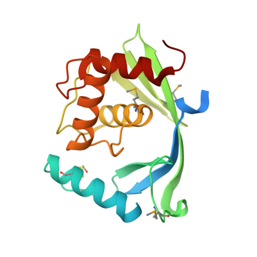
Clostridioides difficile causes life-threatening gastrointestinal infections. It is a high-risk pathogen due to a lack of effective treatments, antimicrobial resistance, and a poorly conserved genomic core. Herein, we report 30 X-ray structures from a structure genomics pipeline spanning 13 years, representing 10.2% of the X-ray structures for this important pathogen.
- Department of Microbiology-Immunology, Feinberg School of Medicine, Northwestern University, Chicago, Illinois, USA.
Organizational Affiliation: