Learning from oligosaccharide soaks of crystals of an AA13 lytic polysaccharide monooxygenase: crystal packing, ligand binding and active-site disorder.
Frandsen, K.E., Poulsen, J.C., Tovborg, M., Johansen, K.S., Lo Leggio, L.(2017) Acta Crystallogr D Struct Biol 73: 64-76
- PubMed: 28045386 Search on PubMed
- DOI: https://doi.org/10.1107/S2059798316019641
- Primary Citation Related Structures:
5LSV, 5T7J, 5T7K, 5T7N - PubMed Abstract:
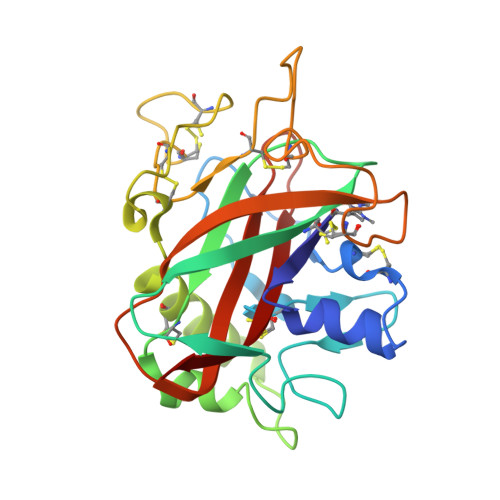
Lytic polysaccharide monooxygenases (LPMOs) are a class of copper-dependent enzymes discovered within the last ten years. They oxidatively cleave polysaccharides (chitin, lignocellulose, hemicellulose and starch-derived), presumably making recalcitrant substrates accessible to glycoside hydrolases. Recently, the first crystal structure of an LPMO-substrate complex was reported, giving insights into the interaction of LPMOs with β-linked substrates (Frandsen et al., 2016). The LPMOs acting on α-linked glycosidic bonds (family AA13) display binding surfaces that are quite different from those of LPMOs that act on β-linked glycosidic bonds (families AA9-AA11), as revealed from the first determined structure (Lo Leggio et al., 2015), and thus presumably the AA13s interact with their substrate in a distinct fashion. Here, several new structures of the same AA13 enzyme, Aspergillus oryzae AA13, are presented. Crystals obtained in the presence of high zinc-ion concentrations were used, as they can be obtained more reproducibly than those used to refine the deposited copper-containing structure. One structure with an ordered zinc-bound active site was solved at 1.65 Å resolution, and three structures from crystals soaked with maltooligosaccharides in solutions devoid of zinc ions were solved at resolutions of up to 1.10 Å. Despite similar unit-cell parameters, small rearrangements in the crystal packing occur when the crystals are depleted of zinc ions, resulting in a more occluded substrate-binding surface. In two of the three structures maltooligosaccharide ligands are bound, but not at the active site. Two of the structures presented show a His-ligand conformation that is incompatible with metal-ion binding. In one of these structures this conformation is the principal one (80% occupancy), giving a rare atomic resolution view of a substantially misfolded enzyme that is presumably rendered inactive.
- Department of Chemistry, University of Copenhagen, Universitetsparken 5, 2100 Copenhagen, Denmark.
Organizational Affiliation: