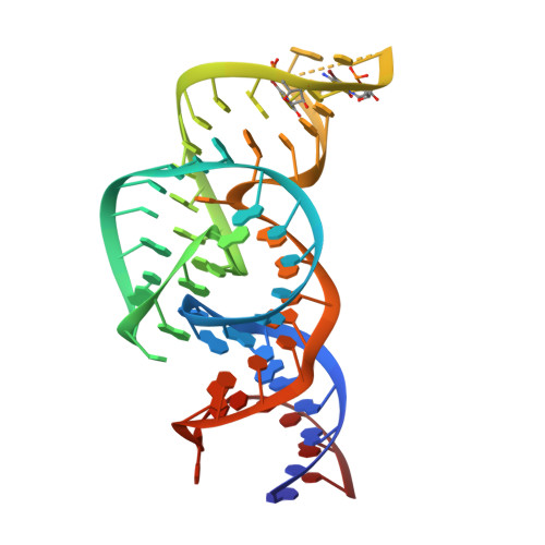
5T5A

Crystal Structure of the Twister Sister (TS) Ribozyme at 2.0 Angstrom


Experimental Data Snapshot

  • Method: X-RAY DIFFRACTION
  • Resolution: 2.00 Å
  • R-Value Free: 0.217 
  • R-Value Work: 0.170 
  • R-Value Observed: 0.172 

wwPDB Validation   3D Report Full Report


This is version 1.3 of the entry. See complete history


Literature

The structure of a nucleolytic ribozyme that employs a catalytic metal ion.

Liu, Y.Wilson, T.J.Lilley, D.M.

(2017) Nat Chem Biol 13: 508-513

  • DOI: https://doi.org/10.1038/nchembio.2333
  • Primary Citation of Related Structures:  
    5T5A

  • PubMed Abstract: 

    The TS ribozyme (originally called "twister sister") is a catalytic RNA. We present a crystal structure of the ribozyme in a pre-reactive conformation. Two co-axial helical stacks are organized by a three-way junction and two tertiary contacts. Five divalent metal ions are directly coordinated to RNA ligands, making important contributions to the RNA architecture. The scissile phosphate lies in a quasihelical loop region that is organized by a network of hydrogen bonding. A divalent metal ion is directly bound to the nucleobase 5' to the scissile phosphate, with an inner-sphere water molecule positioned to interact with the O2' nucleophile. The rate of ribozyme cleavage correlated in a log-linear manner with divalent metal ion pK a , consistent with proton transfer in the transition state, and we propose that the bound metal ion is a likely general base for the cleavage reaction. Our data indicate that the TS ribozyme functions predominantly as a metalloenzyme.


  • Organizational Affiliation

    Cancer Research UK Nucleic Acid Structure Research Group, MSI/WTB Complex, The University of Dundee, Dundee, UK.


Macromolecules
Find similar nucleic acids by: 3D Structure
Entity ID: 1
MoleculeChains LengthOrganismImage
DNA/RNA (71-MER)62metagenome
Sequence Annotations
Expand
  • Reference Sequence
Experimental Data & Validation

Experimental Data

  • Method: X-RAY DIFFRACTION
  • Resolution: 2.00 Å
  • R-Value Free: 0.217 
  • R-Value Work: 0.170 
  • R-Value Observed: 0.172 
  • Space Group: P 41 21 2
Unit Cell:
Length ( Å )Angle ( ˚ )
a = 39.304α = 90
b = 39.304β = 90
c = 228.413γ = 90
Software Package:
Software NamePurpose
PHENIXrefinement
XDSdata reduction
XDSdata scaling
PHENIXphasing

Structure Validation

View Full Validation Report


Entry History & Funding Information

Deposition Data


Funding OrganizationLocationGrant Number
Cancer Research UKUnited Kingdom--

Revision History  (Full details and data files)

  • Version 1.0: 2017-03-08
    Type: Initial release
  • Version 1.1: 2017-03-15
    Changes: Database references
  • Version 1.2: 2017-04-26
    Changes: Database references
  • Version 1.3: 2024-05-08
    Changes: Data collection, Database references, Derived calculations