7-Phenoxy-Substituted 3,4-Dihydro-2H-1,2,4-benzothiadiazine 1,1-Dioxides as Positive Allosteric Modulators of alpha-Amino-3-hydroxy-5-methyl-4-isoxazolepropionic Acid (AMPA) Receptors with Nanomolar Potency.
Goffin, E., Drapier, T., Larsen, A.P., Geubelle, P., Ptak, C.P., Laulumaa, S., Rovinskaja, K., Gilissen, J., Tullio, P., Olsen, L., Frydenvang, K., Pirotte, B., Hanson, J., Oswald, R.E., Kastrup, J.S., Francotte, P.(2018) J Med Chem 61: 251-264
- PubMed: 29256599 Search on PubMedSearch on PubMed Central
- DOI: https://doi.org/10.1021/acs.jmedchem.7b01323
- Primary Citation Related Structures:
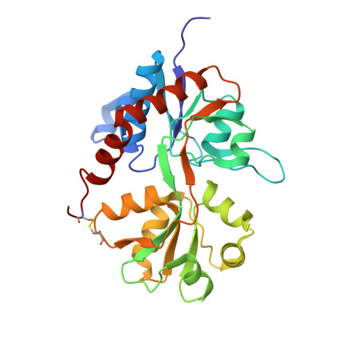
5O9A, 5OEW - PubMed Abstract:
We report here the synthesis of 7-phenoxy-substituted 3,4-dihydro-2H-1,2,4-benzothiadiazine 1,1-dioxides and their evaluation as AMPA receptor positive allosteric modulators (AMPApams). The impact of substitution on the phenoxy ring and on the nitrogen atom at the 4-position was examined. At GluA2(Q) expressed in HEK293 cells (calcium flux experiment), the most potent compound was 11m (4-cyclopropyl-7-(3-methoxyphenoxy)-3,4-dihydro-2H-1,2,4-benzothiadiazine 1,1-dioxide, EC 50 = 2.0 nM). The Hill coefficient in the screening and the shape of the dimerization curve in small-angle X-ray scattering (SAXS) experiments using isolated GluA2 ligand-binding domain (GluA2-LBD) are consistent with binding of one molecule of 11m per dimer interface, contrary to most benzothiadiazine dioxides developed to date. This observation was confirmed by the X-ray structure of 11m bound to GluA2-LBD and by NMR. This is the first benzothiadiazine dioxide AMPApam to reach the nanomolar range.
- Laboratory of Medicinal Chemistry, Center for Interdisciplinary Research on Medicines (CIRM), University of Liège , Quartier Hôpital B36 Av. Hippocrate 15 B-4000 Liège, Belgium.
Organizational Affiliation: