Structural remodeling and oligomerization of human cathelicidin on membranes suggest fibril-like structures as active species.
Sancho-Vaello, E., Francois, P., Bonetti, E.J., Lilie, H., Finger, S., Gil-Ortiz, F., Gil-Carton, D., Zeth, K.(2017) Sci Rep 7: 15371-15371
- PubMed: 29133814 Search on PubMedSearch on PubMed Central
- DOI: https://doi.org/10.1038/s41598-017-14206-1
- Primary Citation Related Structures:
5NMN, 5NNK, 5NNM, 5NNT - PubMed Abstract:
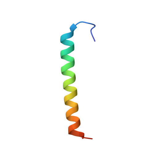
Antimicrobial peptides as part of the mammalian innate immune system target and remove major bacterial pathogens, often through irreversible damage of their cellular membranes. To explore the mechanism by which the important cathelicidin peptide LL-37 of the human innate immune system interacts with membranes, we performed biochemical, biophysical and structural studies. The crystal structure of LL-37 displays dimers of anti-parallel helices and the formation of amphipathic surfaces. Peptide-detergent interactions introduce remodeling of this structure after occupation of defined hydrophobic sites at the dimer interface. Furthermore, hydrophobic nests are shaped between dimer structures providing another scaffold enclosing detergents. Both scaffolds underline the potential of LL-37 to form defined peptide-lipid complexes in vivo. After adopting the activated peptide conformation LL-37 can polymerize and selectively extract bacterial lipids whereby the membrane is destabilized. The supramolecular fibril-like architectures formed in crystals can be reproduced in a peptide-lipid system after nanogold-labelled LL-37 interacted with lipid vesicles as followed by electron microscopy. We suggest that these supramolecular structures represent the LL-37-membrane active state. Collectively, our study provides new insights into the fascinating plasticity of LL-37 demonstrated at atomic resolution and opens the venue for LL-37-based molecules as novel antibiotics.
- Unidad de Biofisica, Centro Mixto Consejo Superior de Investigaciones Científicas-Universidad del País Vasco/Euskal Herriko Unibertsitatea (CSIC,UPV/EHU), Barrio Sarriena s/n, Leioa, Bizkaia, Spain.
Organizational Affiliation: