5NJ8 | pdb_00005nj8

Structural basis for aryl hydrocarbon receptor mediated gene activation


Experimental Data Snapshot

  • Method: X-RAY DIFFRACTION
  • Resolution: 3.30 Å
  • R-Value Free: 
    0.333 (Depositor), 0.338 (DCC) 
  • R-Value Work: 
    0.292 (Depositor), 0.294 (DCC) 
  • R-Value Observed: 
    0.294 (Depositor) 

wwPDB Validation 3D Report Full Report

Validation slider image for 5NJ8

This is version 2.1 of the entry. See complete history

Literature

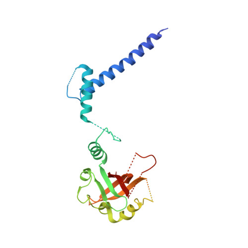
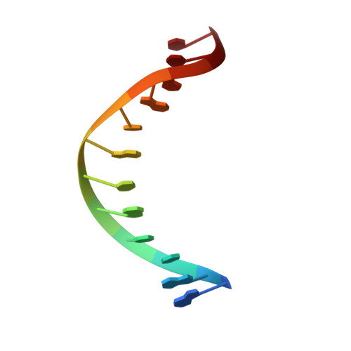
Structural Basis for Aryl Hydrocarbon Receptor-Mediated Gene Activation.

Schulte, K.W.Green, E.Wilz, A.Platten, M.Daumke, O.

(2017) Structure 25: 1025-1033.e3

  • DOI: https://doi.org/10.1016/j.str.2017.05.008
  • Primary Citation Related Structures: 
    5NJ8

  • PubMed Abstract: 

    The aryl hydrocarbon receptor (AHR) and the AHR nuclear translocator (ARNT) constitute a heterodimeric basic helix-loop-helix-Per-ARNT-Sim (bHLH-PAS) domain containing transcription factor with central functions in development and cellular homeostasis. AHR is activated by xenobiotics, notably dioxin, as well as by exogenous and endogenous metabolites. Modulation of AHR activity holds promise for the treatment of diseases featuring altered cellular homeostasis, such as cancer or autoimmune disorders. Here, we present the crystal structure of a heterodimeric AHR:ARNT complex containing the PAS A and bHLH domain bound to its target DNA. The structure provides insights into the DNA binding mode of AHR and elucidates how stable dimerization of AHR:ARNT is achieved through sophisticated domain interplay via three specific interfaces. Using mutational analyses, we prove the relevance of the observed interfaces for AHR-mediated gene activation. Thus, our work establishes the structural basis of AHR assembly and DNA interaction and provides a template for targeted drug design.


  • Organizational Affiliation
    • Crystallography Department, Max-Delbrück-Center for Molecular Medicine, Robert-Rössle-Strasse 10, 13125 Berlin, Germany; Institute of Chemistry and Biochemistry, Freie Universität Berlin, Takustrasse 6, 14195 Berlin, Germany.

Macromolecule Content 

  • Total Structure Weight: 126.5 kDa 
  • Atom Count: 5,848 
  • Modeled Residue Count: 708 
  • Deposited Residue Count: 1,034 
  • Unique protein chains: 2
  • Unique nucleic acid chains: 2

Macromolecules


Find similar proteins by:|  3D Structure
Entity ID: 1
MoleculeChains  Sequence LengthOrganismDetailsImage
Aryl hydrocarbon receptor
A, C
254Homo sapiensMutation(s): 0 
Gene Names: AHRBHLHE76
UniProt & NIH Common Fund Data Resources
Find proteins for P35869 (Homo sapiens)
Explore P35869 
Go to UniProtKB:  P35869
PHAROS:  P35869
GTEx:  ENSG00000106546 
Entity Groups
Sequence Clusters30% Identity50% Identity70% Identity90% Identity95% Identity100% Identity
UniProt GroupP35869
Sequence Annotations
Expand
Reference Sequence
Find similar proteins by:|  3D Structure
Entity ID: 2
MoleculeChains  Sequence LengthOrganismDetailsImage
Aryl hydrocarbon receptor nuclear translocator
B, D
239Mus musculusMutation(s): 1 
Gene Names: Arnt
UniProt & NIH Common Fund Data Resources
Find proteins for P27540 (Homo sapiens)
Explore P27540 
Go to UniProtKB:  P27540
PHAROS:  P27540
GTEx:  ENSG00000143437 
Entity Groups
Sequence Clusters30% Identity50% Identity70% Identity90% Identity95% Identity100% Identity
UniProt GroupP27540
Sequence Annotations
Expand
Reference Sequence
Find similar nucleic acids by:  Sequence
Entity ID: 3
MoleculeChains LengthOrganismImage
DNA (5'-D(*GP*GP*TP*CP*AP*CP*GP*CP*AP*AP*CP*C)-3')
E, G
12Homo sapiens
Sequence Annotations
Expand
Reference Sequence
Find similar nucleic acids by:  Sequence
Entity ID: 4
MoleculeChains LengthOrganismImage
DNA (5'-D(*GP*GP*TP*TP*GP*CP*GP*TP*GP*AP*CP*C)-3')
F, H
12Homo sapiens
Sequence Annotations
Expand
Reference Sequence

Small Molecules

Ligands 2 Unique
IDChains Name / Formula / InChI Key2D Diagram3D Interactions
ER3

Query on ER3


Download:Ideal Coordinates CCD File
I [auth A]
J [auth A]
L [auth A]
M [auth B]
N [auth B]
I [auth A],
J [auth A],
L [auth A],
M [auth B],
N [auth B],
O [auth B],
P [auth B],
Q [auth C],
R [auth C],
S [auth D],
T [auth D],
U [auth H]
ERBIUM (III) ION
Er
JHFPQYFEJICGKC-UHFFFAOYSA-N
ACT

Query on ACT


Download:Ideal Coordinates CCD File
K [auth A]ACETATE ION
C2 H3 O2
QTBSBXVTEAMEQO-UHFFFAOYSA-M

Experimental Data & Validation

Experimental Data

  • Method: X-RAY DIFFRACTION
  • Resolution: 3.30 Å
  • R-Value Free:  0.333 (Depositor), 0.338 (DCC) 
  • R-Value Work:  0.292 (Depositor), 0.294 (DCC) 
  • R-Value Observed: 0.294 (Depositor) 
Space Group: P 65 2 2
Unit Cell:
Length ( Å )Angle ( ˚ )
a = 91.321α = 90
b = 91.321β = 90
c = 464.914γ = 120
Software Package:
Software NamePurpose
PHENIXrefinement
XDSdata reduction
XDSdata scaling
AutoSolphasing

Structure Validation

View Full Validation Report


Entry History 

Deposition Data

Revision History  (Full details and data files)

  • Version 1.0: 2017-06-28
    Type: Initial release
  • Version 1.1: 2017-07-19
    Changes: Database references
  • Version 2.0: 2020-02-05
    Changes: Advisory, Atomic model, Data collection, Database references, Derived calculations, Polymer sequence, Source and taxonomy, Structure summary
  • Version 2.1: 2024-05-08
    Changes: Data collection, Database references, Derived calculations