5MES | pdb_00005mes

MCL1 FAB COMPLEX IN COMPLEX WITH COMPOUND 29


Experimental Data Snapshot

  • Method: X-RAY DIFFRACTION
  • Resolution: 2.24 Å
  • R-Value Free: 
    0.230 (Depositor), 0.218 (DCC) 
  • R-Value Work: 
    0.180 (Depositor) 
  • R-Value Observed: 
    0.182 (Depositor) 

wwPDB Validation 3D Report Full Report

Validation slider image for 5MES

Ligand Structure Quality Assessment 


This is version 1.3 of the entry. See complete history

Literature

Structure Based Design of Non-Natural Peptidic Macrocyclic Mcl-1 Inhibitors.

Johannes, J.W.Bates, S.Beigie, C.Belmonte, M.A.Breen, J.Cao, S.Centrella, P.A.Clark, M.A.Cuozzo, J.W.Dumelin, C.E.Ferguson, A.D.Habeshian, S.Hargreaves, D.Joubran, C.Kazmirski, S.Keefe, A.D.Lamb, M.L.Lan, H.Li, Y.Ma, H.Mlynarski, S.Packer, M.J.Rawlins, P.B.Robbins, D.W.Shen, H.Sigel, E.A.Soutter, H.H.Su, N.Troast, D.M.Wang, H.Wickson, K.F.Wu, C.Zhang, Y.Zhao, Q.Zheng, X.Hird, A.W.

(2017) ACS Med Chem Lett 8: 239-244

  • DOI: https://doi.org/10.1021/acsmedchemlett.6b00464
  • Primary Citation Related Structures: 
    5KU9, 5MES, 5MEV

  • PubMed Abstract: 

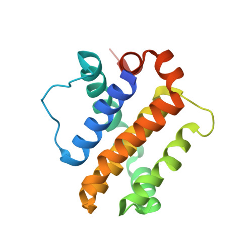
    Mcl-1 is a pro-apoptotic BH3 protein family member similar to Bcl-2 and Bcl-xL. Overexpression of Mcl-1 is often seen in various tumors and allows cancer cells to evade apoptosis. Here we report the discovery and optimization of a series of non-natural peptide Mcl-1 inhibitors. Screening of DNA-encoded libraries resulted in hit compound 1 , a 1.5 μM Mcl-1 inhibitor. A subsequent crystal structure demonstrated that compound 1 bound to Mcl-1 in a β-turn conformation, such that the two ends of the peptide were close together. This proximity allowed for the linking of the two ends of the peptide to form a macrocycle. Macrocyclization resulted in an approximately 10-fold improvement in binding potency. Further exploration of a key hydrophobic interaction with Mcl-1 protein and also with the moiety that engages Arg256 led to additional potency improvements. The use of protein-ligand crystal structures and binding kinetics contributed to the design and understanding of the potency gains. Optimized compound 26 is a <3 nM Mcl-1 inhibitor, while inhibiting Bcl-2 at only 5 μM and Bcl-xL at >99 μM, and induces cleaved caspase-3 in MV4-11 cells with an IC 50 of 3 μM after 6 h.


  • Organizational Affiliation
    • AstraZeneca R&D Boston , 35 Gatehouse Drive, Waltham, Massachusetts 02451, United States.

Macromolecule Content 

  • Total Structure Weight: 67.66 kDa 
  • Atom Count: 4,679 
  • Modeled Residue Count: 577 
  • Deposited Residue Count: 618 
  • Unique protein chains: 3

Macromolecules

Find similar proteins by:|  3D Structure
Entity ID: 1
MoleculeChains  Sequence LengthOrganismDetailsImage
Induced myeloid leukemia cell differentiation protein Mcl-1 homolog,Induced myeloid leukemia cell differentiation protein Mcl-1162Mus musculusHomo sapiens
This entity is chimeric
Mutation(s): 0 
Gene Names: Mcl1MCL1BCL2L3
UniProt & NIH Common Fund Data Resources
Find proteins for P97287 (Mus musculus)
Explore P97287 
Go to UniProtKB:  P97287
IMPC:  MGI:101769
Find proteins for Q07820 (Homo sapiens)
Explore Q07820 
Go to UniProtKB:  Q07820
PHAROS:  Q07820
GTEx:  ENSG00000143384 
Entity Groups
Sequence Clusters30% Identity50% Identity70% Identity90% Identity95% Identity100% Identity
UniProt GroupsP97287Q07820
Sequence Annotations
Expand
Reference Sequence
Find similar proteins by:|  3D Structure
Entity ID: 2
MoleculeChains  Sequence LengthOrganismDetailsImage
Heavy ChainB [auth H]230Homo sapiensMutation(s): 0 
Find similar proteins by:|  3D Structure
Entity ID: 3
MoleculeChains  Sequence LengthOrganismDetailsImage
Light ChainC [auth L]226Homo sapiensMutation(s): 0 

Small Molecules

Ligands 1 Unique
IDChains Name / Formula / InChI Key2D Diagram3D Interactions
7LT

Query on 7LT


Download:Ideal Coordinates CCD File
D [auth A](5~{R},13~{S},17~{S})-5-[[4-chloranyl-3-(2-phenylethyl)phenyl]methyl]-13-[(4-chlorophenyl)methyl]-8-methyl-1,4,8,12,16-pentazatricyclo[15.8.1.0^{20,25}]hexacosa-20(25),21,23-triene-3,7,15,26-tetrone
C44 H49 Cl2 N5 O4
OWZLWLCCVAZXNM-UIJXAYEMSA-N
Binding Affinity Annotations 
IDSourceBinding Affinity
7LT BindingDB:  5MES Kd: 340 (nM) from 1 assay(s)

Experimental Data & Validation

Experimental Data

  • Method: X-RAY DIFFRACTION
  • Resolution: 2.24 Å
  • R-Value Free:  0.230 (Depositor), 0.218 (DCC) 
  • R-Value Work:  0.180 (Depositor) 
  • R-Value Observed: 0.182 (Depositor) 
Space Group: C 1 2 1
Unit Cell:
Length ( Å )Angle ( ˚ )
a = 168.47α = 90
b = 40.33β = 126.11
c = 102.84γ = 90
Software Package:
Software NamePurpose
BUSTERrefinement
MOSFLMdata reduction
SCALAdata scaling
PHASERphasing

Structure Validation

View Full Validation Report


Ligand Structure Quality Assessment 


Entry History 

Deposition Data

Revision History  (Full details and data files)

  • Version 1.0: 2017-01-18
    Type: Initial release
  • Version 1.1: 2017-03-01
    Changes: Database references
  • Version 1.2: 2017-03-08
    Changes: Database references
  • Version 1.3: 2024-10-23
    Changes: Data collection, Database references, Structure summary