5M7R | pdb_00005m7r

Structure of human O-GlcNAc hydrolase


Experimental Data Snapshot

  • Method: X-RAY DIFFRACTION
  • Resolution: 2.35 Å
  • R-Value Free: 
    0.249 (Depositor), 0.250 (DCC) 
  • R-Value Work: 
    0.216 (Depositor), 0.219 (DCC) 
  • R-Value Observed: 
    0.217 (Depositor) 

Starting Model: experimental
View more details

wwPDB Validation 3D Report Full Report

Validation slider image for 5M7R

This is version 1.4 of the entry. See complete history

Literature

Structural and functional insight into human O-GlcNAcase.

Roth, C.Chan, S.Offen, W.A.Hemsworth, G.R.Willems, L.I.King, D.T.Varghese, V.Britton, R.Vocadlo, D.J.Davies, G.J.

(2017) Nat Chem Biol 13: 610-612

  • DOI: https://doi.org/10.1038/nchembio.2358
  • Primary Citation Related Structures: 
    5M7R, 5M7S, 5M7T, 5M7U

  • PubMed Abstract: 

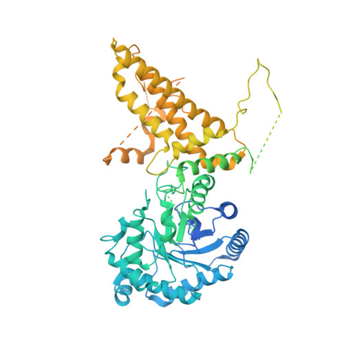
    O-GlcNAc hydrolase (OGA) removes O-linked N-acetylglucosamine (O-GlcNAc) from a myriad of nucleocytoplasmic proteins. Through co-expression and assembly of OGA fragments, we determined the three-dimensional structure of human OGA, revealing an unusual helix-exchanged dimer that lays a structural foundation for an improved understanding of substrate recognition and regulation of OGA. Structures of OGA in complex with a series of inhibitors define a precise blueprint for the design of inhibitors that have clinical value.


  • Organizational Affiliation
    • York Structural Biology Laboratory, Department of Chemistry University of York, York, UK.

Macromolecule Content 

  • Total Structure Weight: 206.04 kDa 
  • Atom Count: 7,704 
  • Modeled Residue Count: 936 
  • Deposited Residue Count: 1,832 
  • Unique protein chains: 1

Macromolecules

Find similar proteins by:|  3D Structure
Entity ID: 1
MoleculeChains  Sequence LengthOrganismDetailsImage
Protein O-GlcNAcase
A, B
916Homo sapiensMutation(s): 0 
Gene Names: MGEA5HEXCKIAA0679MEA5
EC: 3.2.1.169 (PDB Primary Data), 3.2.1 (PDB Primary Data)
UniProt & NIH Common Fund Data Resources
Find proteins for O60502 (Homo sapiens)
Explore O60502 
Go to UniProtKB:  O60502
PHAROS:  O60502
GTEx:  ENSG00000198408 
Entity Groups
Sequence Clusters30% Identity50% Identity70% Identity90% Identity95% Identity100% Identity
UniProt GroupO60502
Sequence Annotations
Expand
Reference Sequence

Experimental Data & Validation

Experimental Data

  • Method: X-RAY DIFFRACTION
  • Resolution: 2.35 Å
  • R-Value Free:  0.249 (Depositor), 0.250 (DCC) 
  • R-Value Work:  0.216 (Depositor), 0.219 (DCC) 
  • R-Value Observed: 0.217 (Depositor) 
Space Group: P 43 21 2
Unit Cell:
Length ( Å )Angle ( ˚ )
a = 102.23α = 90
b = 102.23β = 90
c = 285.49γ = 90
Software Package:
Software NamePurpose
REFMACrefinement
PDB_EXTRACTdata extraction
REFMACrefinement
XDSdata reduction
Aimlessdata scaling
PHASERphasing

Structure Validation

View Full Validation Report


Entry History 

& Funding Information

Deposition Data


Funding OrganizationLocationGrant Number
Biotechnology and Biological Sciences Research CouncilUnited KingdomBB/K003836/1
Canadian Institute of Health ResearchCanadaMOP-123341

Revision History  (Full details and data files)

  • Version 1.0: 2017-03-29
    Type: Initial release
  • Version 1.1: 2017-04-05
    Changes: Database references
  • Version 1.2: 2017-05-24
    Changes: Database references
  • Version 1.3: 2017-08-30
    Changes: Author supporting evidence
  • Version 1.4: 2024-01-17
    Changes: Data collection, Database references, Refinement description