5J6S

Crystal structure of Endoplasmic Reticulum Aminopeptidase 2 (ERAP2) in complex with a hydroxamic derivative ligand


Experimental Data Snapshot

  • Method: X-RAY DIFFRACTION
  • Resolution: 2.80 Å
  • R-Value Free: 0.276 
  • R-Value Work: 0.205 
  • R-Value Observed: 0.208 

wwPDB Validation   3D Report Full Report


Ligand Structure Quality Assessment 


This is version 2.1 of the entry. See complete history


Literature

Crystal Structures of ERAP2 Complexed with Inhibitors Reveal Pharmacophore Requirements for Optimizing Inhibitor Potency.

Mpakali, A.Giastas, P.Deprez-Poulain, R.Papakyriakou, A.Koumantou, D.Gealageas, R.Tsoukalidou, S.Vourloumis, D.Mavridis, I.M.Stratikos, E.Saridakis, E.

(2017) ACS Med Chem Lett 8: 333-337

  • DOI: https://doi.org/10.1021/acsmedchemlett.6b00505
  • Primary Citation of Related Structures:  
    5J6S, 5K1V

  • PubMed Abstract: 

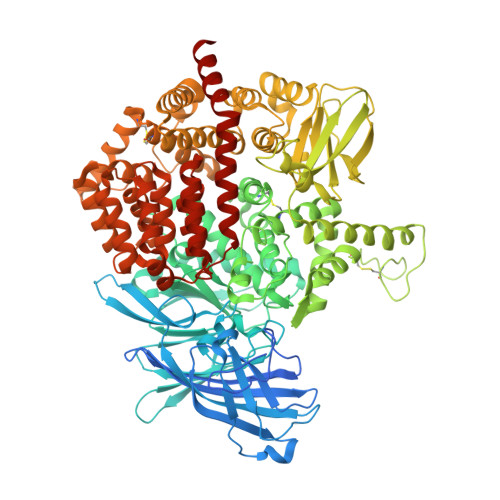
    Endoplasmic reticulum aminopeptidase 2 assists with the generation of antigenic peptides for presentation onto Major Histocompatibility Class I molecules in humans. Recent evidence has suggested that the activity of ERAP2 may contribute to the generation of autoimmunity, thus making ERAP2 a possible pharmacological target for the regulation of adaptive immune responses. To better understand the structural elements of inhibitors that govern their binding affinity to the ERAP2 active site, we cocrystallized ERAP2 with a medium activity 3,4-diaminobenzoic acid inhibitor and a poorly active hydroxamic acid derivative. Comparison of these two crystal structures with a previously solved structure of ERAP2 in complex with a potent phosphinic pseudopeptide inhibitor suggests that engaging the substrate N-terminus recognition properties of the active site is crucial for inhibitor binding even in the absence of a potent zinc-binding group. Proper utilization of all five major pharmacophores is necessary, however, to optimize inhibitor potency.


  • Organizational Affiliation

    National Center for Scientific Research Demokritos , Agia Paraskevi, GR-15310 Athens, Greece.


Macromolecules
Find similar proteins by:  (by identity cutoff)  |  3D Structure
Entity ID: 1
MoleculeChains Sequence LengthOrganismDetailsImage
Endoplasmic reticulum aminopeptidase 2
A, B
967Homo sapiensMutation(s): 1 
Gene Names: ERAP2LRAP
EC: 3.4.11
UniProt & NIH Common Fund Data Resources
Find proteins for Q6P179 (Homo sapiens)
Explore Q6P179 
Go to UniProtKB:  Q6P179
PHAROS:  Q6P179
GTEx:  ENSG00000164308 
Entity Groups  
Sequence Clusters30% Identity50% Identity70% Identity90% Identity95% Identity100% Identity
UniProt GroupQ6P179
Sequence Annotations
Expand
  • Reference Sequence
Oligosaccharides

Help

Entity ID: 2
MoleculeChains Length2D Diagram Glycosylation3D Interactions
2-acetamido-2-deoxy-beta-D-glucopyranose-(1-4)-2-acetamido-2-deoxy-beta-D-glucopyranose
C, E
2N-Glycosylation
Glycosylation Resources
GlyTouCan:  G42666HT
GlyCosmos:  G42666HT
GlyGen:  G42666HT
Entity ID: 3
MoleculeChains Length2D Diagram Glycosylation3D Interactions
beta-D-mannopyranose-(1-4)-2-acetamido-2-deoxy-beta-D-glucopyranose-(1-4)-2-acetamido-2-deoxy-beta-D-glucopyranose
D, G
3N-Glycosylation
Glycosylation Resources
GlyTouCan:  G15407YE
GlyCosmos:  G15407YE
GlyGen:  G15407YE
Entity ID: 4
MoleculeChains Length2D Diagram Glycosylation3D Interactions
alpha-D-mannopyranose-(1-3)-[alpha-D-mannopyranose-(1-6)]beta-D-mannopyranose-(1-4)-2-acetamido-2-deoxy-beta-D-glucopyranose-(1-4)-2-acetamido-2-deoxy-beta-D-glucopyranose
F
5N-Glycosylation
Glycosylation Resources
GlyTouCan:  G22768VO
GlyCosmos:  G22768VO
GlyGen:  G22768VO
Small Molecules
Ligands 3 Unique
IDChains Name / Formula / InChI Key2D Diagram3D Interactions
6GA
Query on 6GA

Download Ideal Coordinates CCD File 
O [auth A],
V [auth B]
(2S)-N~1~-benzyl-2-[(4-fluorophenyl)methyl]-N~3~-hydroxypropanediamide
C17 H17 F N2 O3
UBULRDFJXZRBJH-HNNXBMFYSA-N
NAG
Query on NAG

Download Ideal Coordinates CCD File 
I [auth A]
J [auth A]
K [auth A]
L [auth A]
M [auth A]
I [auth A],
J [auth A],
K [auth A],
L [auth A],
M [auth A],
N [auth A],
Q [auth B],
R [auth B],
S [auth B],
T [auth B],
U [auth B]
2-acetamido-2-deoxy-beta-D-glucopyranose
C8 H15 N O6
OVRNDRQMDRJTHS-FMDGEEDCSA-N
ZN
Query on ZN

Download Ideal Coordinates CCD File 
H [auth A],
P [auth B]
ZINC ION
Zn
PTFCDOFLOPIGGS-UHFFFAOYSA-N
Binding Affinity Annotations 
IDSourceBinding Affinity
6GA BindingDB:  5J6S IC50: 1.00e+5 (nM) from 1 assay(s)
Experimental Data & Validation

Experimental Data

  • Method: X-RAY DIFFRACTION
  • Resolution: 2.80 Å
  • R-Value Free: 0.276 
  • R-Value Work: 0.205 
  • R-Value Observed: 0.208 
  • Space Group: P 1 21 1
Unit Cell:
Length ( Å )Angle ( ˚ )
a = 73.54α = 90
b = 134.33β = 91.71
c = 127.58γ = 90
Software Package:
Software NamePurpose
PHENIXrefinement
iMOSFLMdata reduction
SCALAdata scaling
PHASERphasing

Structure Validation

View Full Validation Report


Ligand Structure Quality Assessment 


Entry History & Funding Information

Deposition Data


Funding OrganizationLocationGrant Number
European Union and the Greek Secretariat for Research and Technology (Bilateral Greece-France collaboration action)GreeceHIAP

Revision History  (Full details and data files)

  • Version 1.0: 2017-03-15
    Type: Initial release
  • Version 1.1: 2017-04-05
    Changes: Database references
  • Version 2.0: 2020-07-29
    Type: Remediation
    Reason: Carbohydrate remediation
    Changes: Advisory, Atomic model, Data collection, Derived calculations, Structure summary
  • Version 2.1: 2024-01-10
    Changes: Data collection, Database references, Derived calculations, Refinement description, Structure summary