5G3T

The structure of the L-tryptophan oxidase VioA from Chromobacterium violaceum


Experimental Data Snapshot

  • Method: X-RAY DIFFRACTION
  • Resolution: 1.80 Å
  • R-Value Free: 0.192 
  • R-Value Work: 0.157 
  • R-Value Observed: 0.159 

wwPDB Validation   3D Report Full Report


Ligand Structure Quality Assessment 


This is version 2.0 of the entry. See complete history


Literature

Biosynthesis of Violacein: Structure and Function of L-Tryptophan Oxidase Vioa Chromobacterium Violaceum

Fuller, J.Roepke, R.Krausze, J.Rennhack, K.E.Daniel, N.P.Blankenfeldt, W.Schulz, S.Jahn, D.Moser, J.

(2016) J Biol Chem 291: 20068

  • DOI: https://doi.org/10.1074/jbc.M116.741561
  • Primary Citation of Related Structures:  
    5G3S, 5G3T, 5G3U

  • PubMed Abstract: 

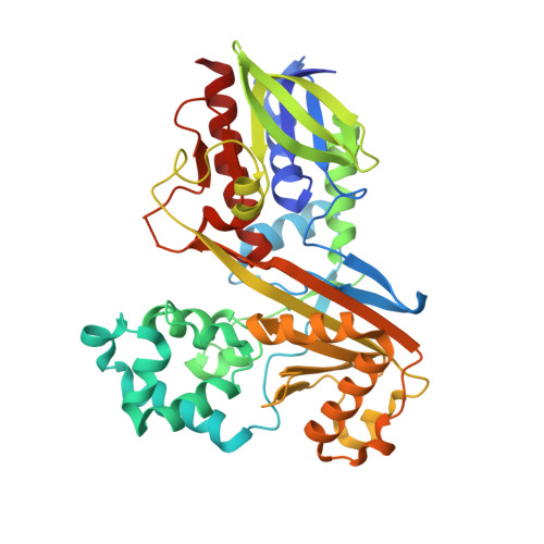
    Violacein is a natural purple pigment of Chromobacterium violaceum with potential medical applications as antimicrobial, antiviral, and anticancer drugs. The initial step of violacein biosynthesis is the oxidative conversion of l-tryptophan into the corresponding α-imine catalyzed by the flavoenzyme l-tryptophan oxidase (VioA). A substrate-related (3-(1H-indol-3-yl)-2-methylpropanoic acid) and a product-related (2-(1H-indol-3-ylmethyl)prop-2-enoic acid) competitive VioA inhibitor was synthesized for subsequent kinetic and x-ray crystallographic investigations. Structures of the binary VioA·FADH2 and of the ternary VioA·FADH2·2-(1H-indol-3-ylmethyl)prop-2-enoic acid complex were resolved. VioA forms a "loosely associated" homodimer as indicated by small-angle x-ray scattering experiments. VioA belongs to the glutathione reductase family 2 of FAD-dependent oxidoreductases according to the structurally conserved cofactor binding domain. The substrate-binding domain of VioA is mainly responsible for the specific recognition of l-tryptophan. Other canonical amino acids were efficiently discriminated with a minor conversion of l-phenylalanine. Furthermore, 7-aza-tryptophan, 1-methyl-tryptophan, 5-methyl-tryptophan, and 5-fluoro-tryptophan were efficient substrates of VioA. The ternary product-related VioA structure indicated involvement of protein domain movement during enzyme catalysis. Extensive structure-based mutagenesis in combination with enzyme kinetics (using l-tryptophan and substrate analogs) identified Arg(64), Lys(269), and Tyr(309) as key catalytic residues of VioA. An increased enzyme activity of protein variant H163A in the presence of l-phenylalanine indicated a functional role of His(163) in substrate binding. The combined structural and mutational analyses lead to the detailed understanding of VioA substrate recognition. Related strategies for the in vivo synthesis of novel violacein derivatives are discussed.


  • Organizational Affiliation

    From the Institute of Microbiology and.


Macromolecules
Find similar proteins by:  (by identity cutoff)  |  3D Structure
Entity ID: 1
MoleculeChains Sequence LengthOrganismDetailsImage
L-TRYPTOPHAN OXIDASE VIOA
A, B, C, D
423Chromobacterium violaceumMutation(s): 1 
EC: 1.4.3.23
UniProt
Find proteins for Q9S3V1 (Chromobacterium violaceum (strain ATCC 12472 / DSM 30191 / JCM 1249 / NBRC 12614 / NCIMB 9131 / NCTC 9757))
Explore Q9S3V1 
Go to UniProtKB:  Q9S3V1
Entity Groups  
Sequence Clusters30% Identity50% Identity70% Identity90% Identity95% Identity100% Identity
UniProt GroupQ9S3V1
Sequence Annotations
Expand
  • Reference Sequence
Small Molecules
Ligands 5 Unique
IDChains Name / Formula / InChI Key2D Diagram3D Interactions
FDA
Query on FDA

Download Ideal Coordinates CCD File 
E [auth A],
I [auth B],
O [auth C],
W [auth D]
DIHYDROFLAVINE-ADENINE DINUCLEOTIDE
C27 H35 N9 O15 P2
YPZRHBJKEMOYQH-UYBVJOGSSA-N
TRS
Query on TRS

Download Ideal Coordinates CCD File 
CA [auth D],
N [auth B],
V [auth C]
2-AMINO-2-HYDROXYMETHYL-PROPANE-1,3-DIOL
C4 H12 N O3
LENZDBCJOHFCAS-UHFFFAOYSA-O
GOL
Query on GOL

Download Ideal Coordinates CCD File 
U [auth C]GLYCEROL
C3 H8 O3
PEDCQBHIVMGVHV-UHFFFAOYSA-N
CL
Query on CL

Download Ideal Coordinates CCD File 
AA [auth D]
BA [auth D]
G [auth A]
H [auth A]
L [auth B]
AA [auth D],
BA [auth D],
G [auth A],
H [auth A],
L [auth B],
M [auth B],
P [auth C],
Q [auth C],
R [auth C],
S [auth C],
T [auth C]
CHLORIDE ION
Cl
VEXZGXHMUGYJMC-UHFFFAOYSA-M
MG
Query on MG

Download Ideal Coordinates CCD File 
F [auth A]
J [auth B]
K [auth B]
X [auth D]
Y [auth D]
F [auth A],
J [auth B],
K [auth B],
X [auth D],
Y [auth D],
Z [auth D]
MAGNESIUM ION
Mg
JLVVSXFLKOJNIY-UHFFFAOYSA-N
Experimental Data & Validation

Experimental Data

  • Method: X-RAY DIFFRACTION
  • Resolution: 1.80 Å
  • R-Value Free: 0.192 
  • R-Value Work: 0.157 
  • R-Value Observed: 0.159 
  • Space Group: P 1 21 1
Unit Cell:
Length ( Å )Angle ( ˚ )
a = 68.09α = 90
b = 89.16β = 92.66
c = 144.43γ = 90
Software Package:
Software NamePurpose
PHENIXrefinement
XDSdata reduction
XSCALEdata scaling
PHASERphasing

Structure Validation

View Full Validation Report


Ligand Structure Quality Assessment 


Entry History 

Deposition Data

Revision History  (Full details and data files)

  • Version 1.0: 2016-08-03
    Type: Initial release
  • Version 1.1: 2016-08-10
    Changes: Database references
  • Version 1.2: 2016-10-05
    Changes: Database references
  • Version 1.3: 2019-03-06
    Changes: Data collection, Experimental preparation, Other
  • Version 2.0: 2019-10-23
    Changes: Atomic model, Data collection, Other