5FJ1

Structure of the standard kink turn HmKt-7 as stem loop in P212121 space group


Experimental Data Snapshot

  • Method: X-RAY DIFFRACTION
  • Resolution: 2.75 Å
  • R-Value Free: 0.183 
  • R-Value Work: 0.167 
  • R-Value Observed: 0.168 

wwPDB Validation   3D Report Full Report


This is version 1.2 of the entry. See complete history


Literature

A Critical Base Pair in K-Turns Determines the Conformational Class Adopted, and Correlates with Biological Function.

Huang, L.Wang, J.Lilley, D.M.J.

(2016) Nucleic Acids Res 44: 5390

  • DOI: https://doi.org/10.1093/nar/gkw201
  • Primary Citation of Related Structures:  
    5FJ0, 5FJ1, 5FJ4, 5FJC, 5FK1, 5FK2, 5FK3, 5FK4, 5FK5, 5FK6, 5FKD, 5FKE, 5FKF, 5FKG, 5FKH

  • PubMed Abstract: 

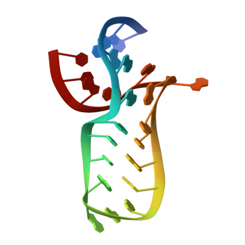
    k-turns are commonly-occurring motifs that introduce sharp kinks into duplex RNA, thereby facilitating tertiary contacts. Both the folding and conformation of k-turns are determined by their local sequence. k-turns fall into two conformational classes, called N3 and N1, that differ in the pattern of hydrogen bonding in the core. We show here that this is determined by the basepair adjacent to the critical G•A pairs. We determined crystal structures of a series of Kt-7 variants in which this 3b,3n position has been systematically varied, showing that this leads to a switch in the conformation. We have previously shown that the 3b,3n position also determines the folding characteristics of the k-turn, i.e. whether or not the k-turn can fold in the presence of metal ions alone. We have analyzed the distribution of 3b,3n sequences from four classes of k-turns from ribosomes, riboswitches and U4 snRNA, finding a strong conservation of properties for a given k-turn type. We thus demonstrate a strong association between biological function, 3b,3n sequence and k-turn folding and conformation. This has strong predictive power, and can be applied to the modeling of large RNA architectures.


  • Organizational Affiliation

    Cancer Research UK Nucleic Acid Structure Research Group, MSI/WTB Complex, The University of Dundee, Dow Street, Dundee DD1 5EH, UK.


Macromolecules

Find similar nucleic acids by:  Sequence   |   3D Structure  

Entity ID: 1
MoleculeChains LengthOrganismImage
HMKT-7
A, B, C, D, E
A, B, C, D, E, F, G, H
24Haloarcula marismortui
Sequence Annotations
Expand
  • Reference Sequence
Small Molecules
Ligands 2 Unique
IDChains Name / Formula / InChI Key2D Diagram3D Interactions
MG
Query on MG

Download Ideal Coordinates CCD File 
AA [auth E]
BA [auth F]
CA [auth F]
DA [auth F]
FA [auth G]
AA [auth E],
BA [auth F],
CA [auth F],
DA [auth F],
FA [auth G],
I [auth A],
J [auth A],
K [auth A],
L [auth A],
O [auth B],
P [auth B],
Q [auth B],
R [auth B],
S [auth B],
T [auth C],
U [auth C],
V [auth C],
W [auth D],
X [auth E],
Y [auth E],
Z [auth E]
MAGNESIUM ION
Mg
JLVVSXFLKOJNIY-UHFFFAOYSA-N
NA
Query on NA

Download Ideal Coordinates CCD File 
EA [auth F],
M [auth A],
N [auth A]
SODIUM ION
Na
FKNQFGJONOIPTF-UHFFFAOYSA-N
Experimental Data & Validation

Experimental Data

  • Method: X-RAY DIFFRACTION
  • Resolution: 2.75 Å
  • R-Value Free: 0.183 
  • R-Value Work: 0.167 
  • R-Value Observed: 0.168 
  • Space Group: P 21 21 21
Unit Cell:
Length ( Å )Angle ( ˚ )
a = 84.052α = 90
b = 84.36β = 90
c = 174.499γ = 90
Software Package:
Software NamePurpose
PHENIXrefinement
iMOSFLMdata reduction
Aimlessdata scaling
MOLREPphasing

Structure Validation

View Full Validation Report


Entry History 

Deposition Data

Revision History  (Full details and data files)

  • Version 1.0: 2016-05-25
    Type: Initial release
  • Version 1.1: 2016-07-06
    Changes: Database references
  • Version 1.2: 2024-01-10
    Changes: Data collection, Database references, Derived calculations, Other, Refinement description