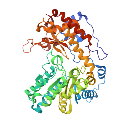
A structural view of the dissociation of Escherichia coli tryptophanase.
Green, K., Qasim, N., Gdaelvsky, G., Kogan, A., Goldgur, Y., Parola, A.H., Lotan, O., Almog, O.(2015) Acta Crystallogr D Biol Crystallogr 71: 2364-2371
- PubMed: 26627645 Search on PubMed
- DOI: https://doi.org/10.1107/S139900471501799X
- Primary Citation Related Structures:
5D8G - PubMed Abstract:
Tryptophanase (Trpase) is a pyridoxal 5'-phosphate (PLP)-dependent homotetrameric enzyme which catalyzes the degradation of L-tryptophan. Trpase is also known for its cold lability, which is a reversible loss of activity at low temperature (2°C) that is associated with the dissociation of the tetramer. Escherichia coli Trpase dissociates into dimers, while Proteus vulgaris Trpase dissociates into monomers. As such, this enzyme is an appropriate model to study the protein-protein interactions and quaternary structure of proteins. The aim of the present study was to understand the differences in the mode of dissociation between the E. coli and P. vulgaris Trpases. In particular, the effect of mutations along the molecular axes of homotetrameric Trpase on its dissociation was studied. To answer this question, two groups of mutants of the E. coli enzyme were created to resemble the amino-acid sequence of P. vulgaris Trpase. In one group, residues 15 and 59 that are located along the molecular axis R (also termed the noncatalytic axis) were mutated. The second group included a mutation at position 298, located along the molecular axis Q (also termed the catalytic axis). Replacing amino-acid residues along the R axis resulted in dissociation of the tetramers into monomers, similar to the P. vulgaris Trpase, while replacing amino-acid residues along the Q axis resulted in dissociation into dimers only. The crystal structure of the V59M mutant of E. coli Trpase was also determined in its apo form and was found to be similar to that of the wild type. This study suggests that in E. coli Trpase hydrophobic interactions along the R axis hold the two monomers together more strongly, preventing the dissociation of the dimers into monomers. Mutation of position 298 along the Q axis to a charged residue resulted in tetramers that are less susceptible to dissociation. Thus, the results indicate that dissociation of E. coli Trpase into dimers occurs along the molecular Q axis.
- Department of Clinical Biochemistry and Pharmacology, Ben-Gurion University of the Negev, PO Box 105, Beer Sheva 84105, Israel.
Organizational Affiliation: