5ZNM | pdb_00005znm

Colicin D Central Domain and C-terminal tRNase domain


Experimental Data Snapshot

  • Method: X-RAY DIFFRACTION
  • Resolution: 1.85 Å
  • R-Value Free: 
    0.212 (Depositor), 0.205 (DCC) 
  • R-Value Work: 
    0.172 (Depositor) 
  • R-Value Observed: 
    0.174 (Depositor) 

wwPDB Validation 3D Report Full Report

Validation slider image for 5ZNM

This is version 1.2 of the entry. See complete history

Literature

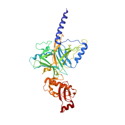
Crystal structure of the central and the C-terminal RNase domains of colicin D implicated its translocation pathway through inner membrane of target cell

Chang, J.W.Sato, Y.Ogawa, T.Arakawa, T.Fukai, S.Fushinobu, S.Masaki, H.

(2018) J Biochem 164: 329-339

  • DOI: https://doi.org/10.1093/jb/mvy056
  • Primary Citation Related Structures: 
    5ZNM

  • PubMed Abstract: 

    Colicins are protein toxins produced by and toxic to Escherichia coli strains. Colicin D consists of an N-terminal domain (NTD), central domain (CD) and C-terminal RNase domain (CRD). The cognate immunity protein, ImmD, is co-synthesized in producer cells to block the toxic tRNase activity of the CRD. Previous studies have reported the crystal structure of CRD/ImmD complex. Colicin D hijacks the surface receptor FepA and the energy transducer TonB system using the NTD for translocation across the outer membrane of the target cells. The CD is required for endoproteolytic processing and the translocation of CRD across the inner membrane, and the membrane-associated protease FtsH and the signal peptidase LepB are exploited in this process. Although several regions of the CD have been identified in interactions with the hijacked inner membrane system or immunity protein, the structural basis of the CD is unknown. In this study, we determined the crystal structure of colicin D, containing both the CD and CRD. The full-length colicin D/ImmD heterodimer structure was built by superimposing the CD-CRD structure with the previously determined partial structures. The overall translocation process of colicin D, including the interaction between CD and LepB, is discussed.


  • Organizational Affiliation
    • Department of Biotechnology, Graduate School of Agricultural and Life Sciences, The University of Tokyo, 1-1-1 Yayoi, Bunkyo-ku, Tokyo, Japan.

Macromolecule Content 

  • Total Structure Weight: 84.9 kDa 
  • Atom Count: 5,805 
  • Modeled Residue Count: 715 
  • Deposited Residue Count: 774 
  • Unique protein chains: 1

Macromolecules

Find similar proteins by:|  3D Structure
Entity ID: 1
MoleculeChains  Sequence LengthOrganismDetailsImage
Colicin-D
A, B
387Escherichia coliMutation(s): 0 
Gene Names: cdacolD
UniProt
Find proteins for P17998 (Escherichia coli)
Explore P17998 
Go to UniProtKB:  P17998
Entity Groups
Sequence Clusters30% Identity50% Identity70% Identity90% Identity95% Identity100% Identity
UniProt GroupP17998
Sequence Annotations
Expand
Reference Sequence

Small Molecules

Ligands 2 Unique
IDChains Name / Formula / InChI Key2D Diagram3D Interactions
SO4

Query on SO4


Download:Ideal Coordinates CCD File
C [auth A]
D [auth A]
E [auth A]
N [auth B]
O [auth B]
C [auth A],
D [auth A],
E [auth A],
N [auth B],
O [auth B],
P [auth B]
SULFATE ION
O4 S
QAOWNCQODCNURD-UHFFFAOYSA-L
GOL

Query on GOL


Download:Ideal Coordinates CCD File
F [auth A]
G [auth A]
H [auth A]
I [auth A]
J [auth A]
F [auth A],
G [auth A],
H [auth A],
I [auth A],
J [auth A],
K [auth A],
L [auth A],
M [auth A],
Q [auth B],
R [auth B],
S [auth B],
T [auth B]
GLYCEROL
C3 H8 O3
PEDCQBHIVMGVHV-UHFFFAOYSA-N

Experimental Data & Validation

Experimental Data

  • Method: X-RAY DIFFRACTION
  • Resolution: 1.85 Å
  • R-Value Free:  0.212 (Depositor), 0.205 (DCC) 
  • R-Value Work:  0.172 (Depositor) 
  • R-Value Observed: 0.174 (Depositor) 
Space Group: C 1 2 1
Unit Cell:
Length ( Å )Angle ( ˚ )
a = 116.352α = 90
b = 146.115β = 102.49
c = 45.763γ = 90
Software Package:
Software NamePurpose
PHENIXrefinement
PHENIXphasing

Structure Validation

View Full Validation Report


Entry History 

& Funding Information

Deposition Data


Funding OrganizationLocationGrant Number
Japan Science and TechnologyJapan17103789

Revision History  (Full details and data files)

  • Version 1.0: 2018-08-29
    Type: Initial release
  • Version 1.1: 2018-10-31
    Changes: Data collection, Database references
  • Version 1.2: 2024-03-27
    Changes: Data collection, Database references