5ZKQ

Crystal structure of the human platelet-activating factor receptor in complex with ABT-491


Experimental Data Snapshot

  • Method: X-RAY DIFFRACTION
  • Resolution: 2.90 Å
  • R-Value Free: 0.235 
  • R-Value Work: 0.203 
  • R-Value Observed: 0.204 

wwPDB Validation   3D Report Full Report


Ligand Structure Quality Assessment 


This is version 1.1 of the entry. See complete history


Literature

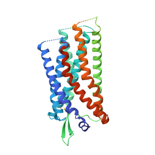
Structural basis for signal recognition and transduction by platelet-activating-factor receptor.

Cao, C.Tan, Q.Xu, C.He, L.Yang, L.Zhou, Y.Zhou, Y.Qiao, A.Lu, M.Yi, C.Han, G.W.Wang, X.Li, X.Yang, H.Rao, Z.Jiang, H.Zhao, Y.Liu, J.Stevens, R.C.Zhao, Q.Zhang, X.C.Wu, B.

(2018) Nat Struct Mol Biol 25: 488-495

  • DOI: https://doi.org/10.1038/s41594-018-0068-y
  • Primary Citation of Related Structures:  
    5ZKP, 5ZKQ

  • PubMed Abstract: 

    Platelet-activating-factor receptor (PAFR) responds to platelet-activating factor (PAF), a phospholipid mediator of cell-to-cell communication that exhibits diverse physiological effects. PAFR is considered an important drug target for treating asthma, inflammation and cardiovascular diseases. Here we report crystal structures of human PAFR in complex with the antagonist SR 27417 and the inverse agonist ABT-491 at 2.8-Å and 2.9-Å resolution, respectively. The structures, supported by molecular docking of PAF, provide insights into the signal-recognition mechanisms of PAFR. The PAFR-SR 27417 structure reveals an unusual conformation showing that the intracellular tips of helices II and IV shift outward by 13 Å and 4 Å, respectively, and helix VIII adopts an inward conformation. The PAFR structures, combined with single-molecule FRET and cell-based functional assays, suggest that the conformational change in the helical bundle is ligand dependent and plays a critical role in PAFR activation, thus greatly extending knowledge about signaling by G-protein-coupled receptors.


  • Organizational Affiliation

    National Laboratory of Biomacromolecules, National Center of Protein Science-Beijing, CAS Center for Excellence in Biomacromolecules, Institute of Biophysics, Chinese Academy of Sciences, Beijing, China.


Macromolecules
Find similar proteins by:  (by identity cutoff)  |  3D Structure
Entity ID: 1
MoleculeChains Sequence LengthOrganismDetailsImage
Platelet-activating factor receptor,Endolysin,Endolysin,Platelet-activating factor receptor
A, B
438Homo sapiensTequatrovirus T4Mutation(s): 7 
Gene Names: PTAFRPAFR
EC: 3.2.1.17
Membrane Entity: Yes 
UniProt & NIH Common Fund Data Resources
Find proteins for P00720 (Enterobacteria phage T4)
Explore P00720 
Go to UniProtKB:  P00720
Find proteins for P25105 (Homo sapiens)
Explore P25105 
Go to UniProtKB:  P25105
PHAROS:  P25105
GTEx:  ENSG00000169403 
Entity Groups  
Sequence Clusters30% Identity50% Identity70% Identity90% Identity95% Identity100% Identity
UniProt GroupsP00720P25105
Sequence Annotations
Expand
  • Reference Sequence
Small Molecules
Ligands 4 Unique
IDChains Name / Formula / InChI Key2D Diagram3D Interactions
9EU
Query on 9EU

Download Ideal Coordinates CCD File 
C [auth A],
I [auth B]
4-ethynyl-3-{3-fluoro-4-[(2-methyl-1H-imidazo[4,5-c]pyridin-1-yl)methyl]benzene-1-carbonyl}-N,N-dimethyl-1H-indole-1-carboxamide
C28 H22 F N5 O2
GDLNHSUSOZEAOR-UHFFFAOYSA-N
OLC
Query on OLC

Download Ideal Coordinates CCD File 
D [auth A]
E [auth A]
F [auth A]
G [auth A]
J [auth B]
D [auth A],
E [auth A],
F [auth A],
G [auth A],
J [auth B],
K [auth B],
L [auth B]
(2R)-2,3-dihydroxypropyl (9Z)-octadec-9-enoate
C21 H40 O4
RZRNAYUHWVFMIP-GDCKJWNLSA-N
SO4
Query on SO4

Download Ideal Coordinates CCD File 
N [auth B]
O [auth B]
P [auth B]
Q [auth B]
R [auth B]
N [auth B],
O [auth B],
P [auth B],
Q [auth B],
R [auth B],
S [auth B]
SULFATE ION
O4 S
QAOWNCQODCNURD-UHFFFAOYSA-L
ZN
Query on ZN

Download Ideal Coordinates CCD File 
H [auth A],
M [auth B]
ZINC ION
Zn
PTFCDOFLOPIGGS-UHFFFAOYSA-N
Experimental Data & Validation

Experimental Data

  • Method: X-RAY DIFFRACTION
  • Resolution: 2.90 Å
  • R-Value Free: 0.235 
  • R-Value Work: 0.203 
  • R-Value Observed: 0.204 
  • Space Group: C 1 2 1
Unit Cell:
Length ( Å )Angle ( ˚ )
a = 86.22α = 90
b = 166.54β = 100
c = 100.31γ = 90
Software Package:
Software NamePurpose
BUSTERrefinement
XDSdata reduction
XDSdata scaling
PHASERphasing

Structure Validation

View Full Validation Report


Ligand Structure Quality Assessment 


Entry History & Funding Information

Deposition Data


Funding OrganizationLocationGrant Number
National Science Foundation (China)China81525024
Ministry of Education (China)ChinaB08029

Revision History  (Full details and data files)

  • Version 1.0: 2018-06-20
    Type: Initial release
  • Version 1.1: 2023-11-22
    Changes: Data collection, Database references, Refinement description