5ZFW

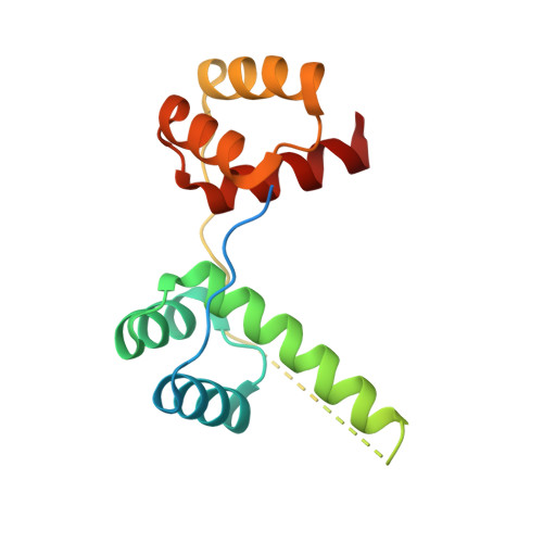
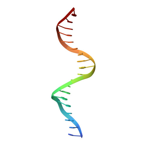
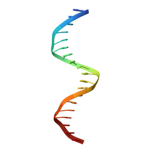
Crystal structure of human DUX4 homeodomains bound to A11G DNA mutant


Experimental Data Snapshot

  • Method: X-RAY DIFFRACTION
  • Resolution: 2.10 Å
  • R-Value Free: 0.227 
  • R-Value Work: 0.192 
  • R-Value Observed: 0.194 

wwPDB Validation   3D Report Full Report


This is version 1.2 of the entry. See complete history


Literature

Structural basis for multiple gene regulation by human DUX4.

Li, Y.Wu, B.Liu, H.Gao, Y.Yang, C.Chen, X.Zhang, J.Chen, Y.Gu, Y.Li, J.Ma, J.Gan, J.

(2018) Biochem Biophys Res Commun 505: 1161-1167

  • DOI: https://doi.org/10.1016/j.bbrc.2018.10.056
  • Primary Citation of Related Structures:  
    5Z6Z, 5ZFW, 5ZFY, 5ZFZ

  • PubMed Abstract: 

    DUX4 plays critical role in the molecular pathogenesis of the neuromuscular disorder facioscapulohumeral muscular dystrophy and acute lymphoblastic leukemia in humans. As a master transcription regulator, DUX4 can also bind the promoters and activate the transcription of hundreds ZGA-associated genes. Here we report on the structural and biochemical studies of DUX4 double homeodomains (DUX4-DH), representing the only structures contain both homeodomain 1 (HD1) and homeodomain 2 (HD2). HD1 and HD2 adopt classical homeobox fold; via the helix inserted into the major groove and the N-terminal extended loop inserted into the minor groove, HD1 and HD2 recognize the box1 (5'-TAA-3') and box2 (5'-TGA-3') nucleotides of the consensus sequence, respectively. Among the box1 and box2 linking nucleotides (CCTAA), the two adenine residues are reported to be highly conserved; however, they are not directly recognized by DUX4-DH in the structures. Besides different nucleotides, our ITC analysis indicated that DUX4-DH can also tolerate various changes in the linker length. Our studies not only revealed the basis for target DNA recognition by DUX4, but also advanced our understanding on multiple gene activation by DUX4.


  • Organizational Affiliation

    State Key Laboratory of Genetic Engineering, Collaborative Innovation Center of Genetics and Development, Department of Physiology and Biophysics, School of Life Sciences, Fudan University, Shanghai, 200438, China.


Macromolecules

Find similar proteins by:  (by identity cutoff)  |  3D Structure
Entity ID: 1
MoleculeChains Sequence LengthOrganismDetailsImage
Double homeobox protein 4-like protein 4149Homo sapiensMutation(s): 0 
Gene Names: DUX4L4
UniProt & NIH Common Fund Data Resources
Find proteins for Q9UBX2 (Homo sapiens)
Explore Q9UBX2 
Go to UniProtKB:  Q9UBX2
PHAROS:  Q9UBX2
GTEx:  ENSG00000260596 
Entity Groups  
Sequence Clusters30% Identity50% Identity70% Identity90% Identity95% Identity100% Identity
UniProt GroupQ9UBX2
Sequence Annotations
Expand
  • Reference Sequence

Find similar nucleic acids by:  Sequence   |   3D Structure  

Entity ID: 2
MoleculeChains LengthOrganismImage
DNA (5'-D(*CP*CP*AP*CP*TP*AP*AP*CP*CP*TP*GP*AP*TP*CP*AP*CP*AP*CP*C)-3')B [auth D]19synthetic construct
Sequence Annotations
Expand
  • Reference Sequence

Find similar nucleic acids by:  Sequence   |   3D Structure  

Entity ID: 3
MoleculeChains LengthOrganismImage
DNA (5'-D(*GP*GP*TP*GP*TP*GP*AP*TP*CP*AP*GP*GP*TP*TP*AP*GP*TP*GP*G)-3')C [auth E]19synthetic construct
Sequence Annotations
Expand
  • Reference Sequence
Experimental Data & Validation

Experimental Data

  • Method: X-RAY DIFFRACTION
  • Resolution: 2.10 Å
  • R-Value Free: 0.227 
  • R-Value Work: 0.192 
  • R-Value Observed: 0.194 
  • Space Group: P 32 2 1
Unit Cell:
Length ( Å )Angle ( ˚ )
a = 64.748α = 90
b = 64.748β = 90
c = 153.484γ = 120
Software Package:
Software NamePurpose
PHENIXrefinement
HKL-3000data reduction
HKL-3000data scaling
PHASERphasing

Structure Validation

View Full Validation Report


Entry History 

Deposition Data

Revision History  (Full details and data files)

  • Version 1.0: 2018-10-31
    Type: Initial release
  • Version 1.1: 2018-11-14
    Changes: Data collection, Database references
  • Version 1.2: 2023-11-22
    Changes: Data collection, Database references, Refinement description