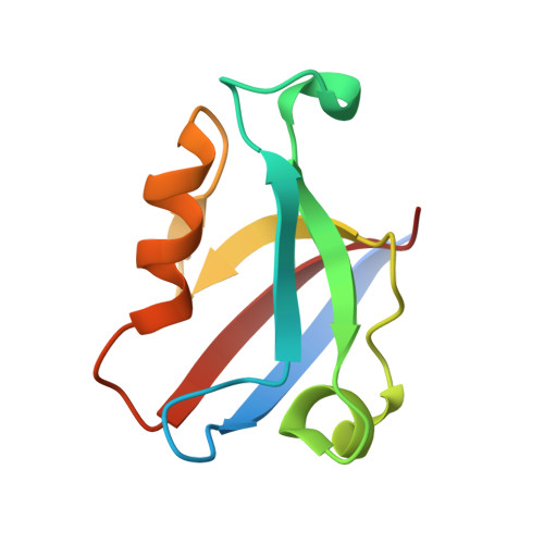
The second PDZ domain of scaffold protein Frmpd2 binds to GluN2A of NMDA receptors.
Lu, X., Zhang, Q., Wang, T.(2019) Biochem Biophys Res Commun 516: 63-67
- PubMed: 31196628
- DOI: https://doi.org/10.1016/j.bbrc.2019.05.087
- Primary Citation of Related Structures:
5ZDS - PubMed Abstract:
The scaffold proteins Frmpd2 is localized at the basolateral membranes of polarized epithelial cells and associated with tight junction formation. In this report, we found that the Frmpd2 is specifically expressed at postsynaptic membrane. By using of co-immunoprecipitation and GST pull-down, Frmpd2 was reported to interact with postsynaptic excitatory N-methyl-d-aspartic acid (NMDA) receptors in vivo and in vitro. In addition, we demonstrated that the second PDZ (PDZ2) domain but not the first or third PDZ domain of Frmpd2 binds to the C-terminus of GluN2A and GluN2B, two subunits of NMDA receptors. By surface plasmon resonance, the affinity of Frmpd2-isolated PDZ2 to GluN2A and GluN2B was identified, which indicates that the interaction of Frmpd2 to GluN2A subunit is more strongly than that to GluN2B subunit. The crystal structure of the PDZ2 domain of the mouse homologue of Frmpd2 was further solved. Some amino acid residues of the PDZ2 structure are supposed to associate with the GluN2A binding. Our study suggests that the scaffold protein Frmpd2 is probably involved in synaptic NMDA receptors-mediated neural excitatory and neurotoxicity in a PDZ2 domain-dependent manner.
- Department of Neurology, Chongqing Key Laboratory of Neurology, The First Affiliated Hospital of Chongqing Medical University, 1 Youyi Road, Chongqing, 400016, China. Electronic address: lxi2646@163.com.
Organizational Affiliation:


