5YRT

Diol dehydratase, AdoCbl/substrate-free, anaerobically-prepared crystal


Experimental Data Snapshot

  • Method: X-RAY DIFFRACTION
  • Resolution: 1.70 Å
  • R-Value Free: 0.187 
  • R-Value Work: 0.149 
  • R-Value Observed: 0.150 

wwPDB Validation   3D Report Full Report


Ligand Structure Quality Assessment 


This is version 1.1 of the entry. See complete history


Literature

Direct Participation of a Peripheral Side Chain of a Corrin Ring in Coenzyme B12Catalysis.

Shibata, N.Sueyoshi, Y.Higuchi, Y.Toraya, T.

(2018) Angew Chem Int Ed Engl 57: 7830-7835

  • DOI: https://doi.org/10.1002/anie.201803591
  • Primary Citation of Related Structures:  
    5YRT, 5YRV, 5YSH, 5YSN, 5YSR

  • PubMed Abstract: 

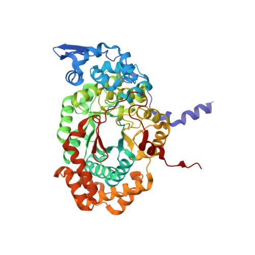
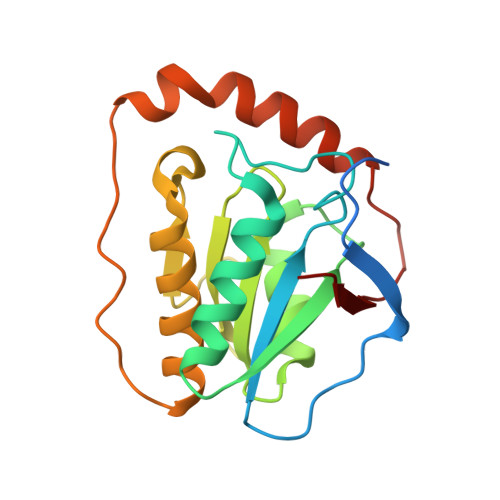
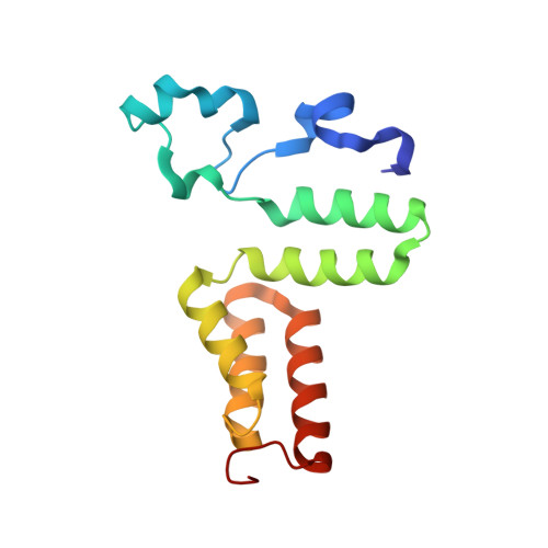
    The crystal structures of the B 12 -dependent isomerases (eliminating) diol dehydratase and ethanolamine ammonia-lyase complexed with adenosylcobalamin were solved with and without substrates. The structures revealed that the peripheral a-acetamide side chain of the corrin ring directly interacts with the adenosyl group to maintain the group in the catalytic position, and that this side chain swings between the original and catalytic positions in a synchronized manner with the radical shuttling between the coenzyme and substrate/product. Mutations involving key residues that cooperatively participate in the positioning of the adenosyl group, directly or indirectly through the interaction with the a-side chain, decreased the turnover rate and increased the relative rate of irreversible inactivation caused by undesirable side reactions. These findings guide the engineering of enzymes for improved catalysis and producing useful chemicals by utilizing the high reactivity of radical species.


  • Organizational Affiliation

    Department of Picobiology/Life Science, Graduate School of Life Science, University of Hyogo, 3-2-1 Koto, Kamigori-cho, Ako-gun, Hyogo, 678-1297, Japan.


Macromolecules
Find similar proteins by:  (by identity cutoff)  |  3D Structure
Entity ID: 1
MoleculeChains Sequence LengthOrganismDetailsImage
Diol dehydrase alpha subunit
A, D, G, J
554Klebsiella oxytocaMutation(s): 0 
Gene Names: pddApduCAB185_12495SAMEA2273575_05741
EC: 4.2.1.28
UniProt
Find proteins for Q59470 (Klebsiella oxytoca)
Explore Q59470 
Go to UniProtKB:  Q59470
Entity Groups  
Sequence Clusters30% Identity50% Identity70% Identity90% Identity95% Identity100% Identity
UniProt GroupQ59470
Sequence Annotations
Expand
  • Reference Sequence
Find similar proteins by:  (by identity cutoff)  |  3D Structure
Entity ID: 2
MoleculeChains Sequence LengthOrganismDetailsImage
Diol dehydrase beta subunit
B, E, H, K
200Klebsiella oxytocaMutation(s): 0 
Gene Names: pddB
EC: 4.2.1.28
UniProt
Find proteins for Q59471 (Klebsiella oxytoca)
Explore Q59471 
Go to UniProtKB:  Q59471
Entity Groups  
Sequence Clusters30% Identity50% Identity70% Identity90% Identity95% Identity100% Identity
UniProt GroupQ59471
Sequence Annotations
Expand
  • Reference Sequence
Find similar proteins by:  (by identity cutoff)  |  3D Structure
Entity ID: 3
MoleculeChains Sequence LengthOrganismDetailsImage
Diol dehydrase gamma subunit
C, F, I, L
137Klebsiella oxytocaMutation(s): 0 
Gene Names: pddCpduEpduE_1AB185_12485SAMEA2273639_01293SAMEA2273697_01477
EC: 4.2.1.28
UniProt
Find proteins for Q59472 (Klebsiella oxytoca)
Explore Q59472 
Go to UniProtKB:  Q59472
Entity Groups  
Sequence Clusters30% Identity50% Identity70% Identity90% Identity95% Identity100% Identity
UniProt GroupQ59472
Sequence Annotations
Expand
  • Reference Sequence
Small Molecules
Ligands 5 Unique
IDChains Name / Formula / InChI Key2D Diagram3D Interactions
B12
Query on B12

Download Ideal Coordinates CCD File 
AA [auth E],
IA [auth H],
QA [auth K],
S [auth B]
COBALAMIN
C62 H89 Co N13 O14 P
LKVIQTCSMMVGFU-DWSMJLPVSA-N
5AD
Query on 5AD

Download Ideal Coordinates CCD File 
FA [auth G],
NA [auth J],
P [auth A],
X [auth D]
5'-DEOXYADENOSINE
C10 H13 N5 O3
XGYIMTFOTBMPFP-KQYNXXCUSA-N
CA
Query on CA

Download Ideal Coordinates CCD File 
CA [auth G],
KA [auth J],
M [auth A],
U [auth D]
CALCIUM ION
Ca
BHPQYMZQTOCNFJ-UHFFFAOYSA-N
K
Query on K

Download Ideal Coordinates CCD File 
DA [auth G]
EA [auth G]
LA [auth J]
MA [auth J]
N [auth A]
DA [auth G],
EA [auth G],
LA [auth J],
MA [auth J],
N [auth A],
O [auth A],
V [auth D],
W [auth D]
POTASSIUM ION
K
NPYPAHLBTDXSSS-UHFFFAOYSA-N
CL
Query on CL

Download Ideal Coordinates CCD File 
BA [auth F]
GA [auth G]
HA [auth G]
JA [auth I]
OA [auth J]
BA [auth F],
GA [auth G],
HA [auth G],
JA [auth I],
OA [auth J],
PA [auth J],
Q [auth A],
R [auth A],
RA [auth L],
T [auth C],
Y [auth D],
Z [auth D]
CHLORIDE ION
Cl
VEXZGXHMUGYJMC-UHFFFAOYSA-M
Experimental Data & Validation

Experimental Data

  • Method: X-RAY DIFFRACTION
  • Resolution: 1.70 Å
  • R-Value Free: 0.187 
  • R-Value Work: 0.149 
  • R-Value Observed: 0.150 
  • Space Group: P 1
Unit Cell:
Length ( Å )Angle ( ˚ )
a = 72.687α = 80.78
b = 95.459β = 87.58
c = 114.567γ = 81.1
Software Package:
Software NamePurpose
PHENIXrefinement
XDSdata reduction
XDSdata scaling
PHASERphasing

Structure Validation

View Full Validation Report


Ligand Structure Quality Assessment 


Entry History 

Deposition Data

  • Released Date: 2018-09-19 
  • Deposition Author(s): Shibata, N.

Revision History  (Full details and data files)

  • Version 1.0: 2018-09-19
    Type: Initial release
  • Version 1.1: 2023-11-22
    Changes: Data collection, Database references, Derived calculations, Refinement description