5YQZ

Structure of the glucagon receptor in complex with a glucagon analogue


Experimental Data Snapshot

  • Method: X-RAY DIFFRACTION
  • Resolution: 3.00 Å
  • R-Value Free: 0.261 
  • R-Value Work: 0.232 
  • R-Value Observed: 0.233 

Starting Models: experimental
View more details

wwPDB Validation   3D Report Full Report


Ligand Structure Quality Assessment 


This is version 2.1 of the entry. See complete history


Literature

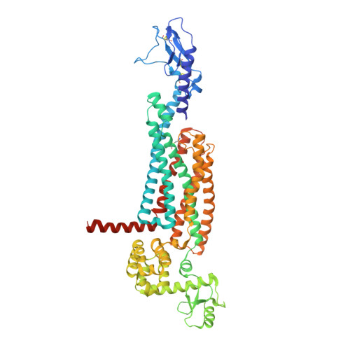
Structure of the glucagon receptor in complex with a glucagon analogue.

Zhang, H.Qiao, A.Yang, L.Van Eps, N.Frederiksen, K.S.Yang, D.Dai, A.Cai, X.Zhang, H.Yi, C.Cao, C.He, L.Yang, H.Lau, J.Ernst, O.P.Hanson, M.A.Stevens, R.C.Wang, M.W.Reedtz-Runge, S.Jiang, H.Zhao, Q.Wu, B.

(2018) Nature 553: 106-110

  • DOI: https://doi.org/10.1038/nature25153
  • Primary Citation of Related Structures:  
    5YQZ

  • PubMed Abstract: 

    Class B G-protein-coupled receptors (GPCRs), which consist of an extracellular domain (ECD) and a transmembrane domain (TMD), respond to secretin peptides to play a key part in hormonal homeostasis, and are important therapeutic targets for a variety of diseases. Previous work has suggested that peptide ligands bind to class B GPCRs according to a two-domain binding model, in which the C-terminal region of the peptide targets the ECD and the N-terminal region of the peptide binds to the TMD binding pocket. Recently, three structures of class B GPCRs in complex with peptide ligands have been solved. These structures provide essential insights into peptide ligand recognition by class B GPCRs. However, owing to resolution limitations, the specific molecular interactions for peptide binding to class B GPCRs remain ambiguous. Moreover, these previously solved structures have different ECD conformations relative to the TMD, which introduces questions regarding inter-domain conformational flexibility and the changes required for receptor activation. Here we report the 3.0 Å-resolution crystal structure of the full-length human glucagon receptor (GCGR) in complex with a glucagon analogue and partial agonist, NNC1702. This structure provides molecular details of the interactions between GCGR and the peptide ligand. It reveals a marked change in the relative orientation between the ECD and TMD of GCGR compared to the previously solved structure of the inactive GCGR-NNC0640-mAb1 complex. Notably, the stalk region and the first extracellular loop undergo major conformational changes in secondary structure during peptide binding, forming key interactions with the peptide. We further propose a dual-binding-site trigger model for GCGR activation-which requires conformational changes of the stalk, first extracellular loop and TMD-that extends our understanding of the previously established two-domain peptide-binding model of class B GPCRs.


  • Organizational Affiliation

    CAS Key Laboratory of Receptor Research, Shanghai Institute of Materia Medica, Chinese Academy of Sciences, 555 Zuchongzhi Road, Pudong, Shanghai 201203, China.


Macromolecules
Find similar proteins by:  (by identity cutoff)  |  3D Structure
Entity ID: 1
MoleculeChains Sequence LengthOrganismDetailsImage
Glucagon receptor,Endolysin,Glucagon receptorA [auth R]575Homo sapiensTequatrovirus T4Mutation(s): 3 
Gene Names: GCGReT4Tp126
EC: 3.2.1.17
Membrane Entity: Yes 
UniProt & NIH Common Fund Data Resources
Find proteins for P47871 (Homo sapiens)
Explore P47871 
Go to UniProtKB:  P47871
GTEx:  ENSG00000215644 
Find proteins for P00720 (Enterobacteria phage T4)
Explore P00720 
Go to UniProtKB:  P00720
Entity Groups  
Sequence Clusters30% Identity50% Identity70% Identity90% Identity95% Identity100% Identity
UniProt GroupsP00720P47871
Glycosylation
Glycosylation Sites: 2Go to GlyGen: P47871-1
Sequence Annotations
Expand
  • Reference Sequence
Find similar proteins by:  (by identity cutoff)  |  3D Structure
Entity ID: 2
MoleculeChains Sequence LengthOrganismDetailsImage
Glucagon analogueB [auth P]28Homo sapiensMutation(s): 0 
Membrane Entity: Yes 
UniProt & NIH Common Fund Data Resources
Find proteins for P01275 (Homo sapiens)
Explore P01275 
Go to UniProtKB:  P01275
GTEx:  ENSG00000115263 
Entity Groups  
Sequence Clusters30% Identity50% Identity70% Identity90% Identity95% Identity100% Identity
UniProt GroupP01275
Sequence Annotations
Expand
  • Reference Sequence
Oligosaccharides

Help

Entity ID: 3
MoleculeChains Length2D Diagram Glycosylation3D Interactions
2-acetamido-2-deoxy-beta-D-glucopyranose-(1-4)-2-acetamido-2-deoxy-beta-D-glucopyranoseC [auth A],
D [auth B]
2N-Glycosylation
Glycosylation Resources
GlyTouCan:  G42666HT
GlyCosmos:  G42666HT
GlyGen:  G42666HT
Experimental Data & Validation

Experimental Data

  • Method: X-RAY DIFFRACTION
  • Resolution: 3.00 Å
  • R-Value Free: 0.261 
  • R-Value Work: 0.232 
  • R-Value Observed: 0.233 
  • Space Group: P 21 21 21
Unit Cell:
Length ( Å )Angle ( ˚ )
a = 60.12α = 90
b = 108.79β = 90
c = 216.25γ = 90
Software Package:
Software NamePurpose
BUSTERrefinement
XDSdata scaling
PHASERphasing

Structure Validation

View Full Validation Report


Ligand Structure Quality Assessment 


Entry History & Funding Information

Deposition Data


Funding OrganizationLocationGrant Number
National Science Foundation (China)China31422017
National Science Foundation (China)China81525024

Revision History  (Full details and data files)

  • Version 1.0: 2018-01-17
    Type: Initial release
  • Version 2.0: 2020-07-29
    Type: Remediation
    Reason: Carbohydrate remediation
    Changes: Advisory, Atomic model, Data collection, Derived calculations, Structure summary
  • Version 2.1: 2023-11-22
    Changes: Data collection, Database references, Refinement description, Structure summary