Structural Basis for pH-Dependent Oligomerization of Dihydropyrimidinase fromPseudomonas aeruginosaPAO1.
Cheng, J.H., Huang, C.C., Huang, Y.H., Huang, C.Y.(2018) Bioinorg Chem Appl 2018: 9564391-9564391
- PubMed: 29666631 Search on PubMedSearch on PubMed Central
- DOI: https://doi.org/10.1155/2018/9564391
- Primary Citation Related Structures:
5YKD - PubMed Abstract:
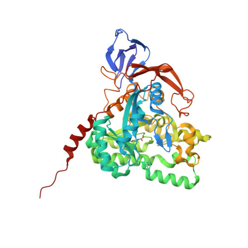
Dihydropyrimidinase, a dimetalloenzyme containing a carboxylated lysine within the active site, is a member of the cyclic amidohydrolase family, which also includes allantoinase, dihydroorotase, hydantoinase, and imidase. Unlike all known dihydropyrimidinases, which are tetrameric, pseudomonal dihydropyrimidinase forms a dimer at neutral pH. In this paper, we report the crystal structure of P. aeruginosa dihydropyrimidinase at pH 5.9 (PDB entry 5YKD). The crystals of P. aeruginosa dihydropyrimidinase belonged to space group C 222 1 with cell dimensions of a = 108.9, b = 155.7, and c = 235.6 Å. The structure of P. aeruginosa dihydropyrimidinase was solved at 2.17 Å resolution. An asymmetric unit of the crystal contained four crystallographically independent P. aeruginosa dihydropyrimidinase monomers. Gel filtration chromatographic analysis of purified P. aeruginosa dihydropyrimidinase revealed a mixture of dimers and tetramers at pH 5.9. Thus, P. aeruginosa dihydropyrimidinase can form a stable tetramer both in the crystalline state and in the solution. Based on sequence analysis and structural comparison of the dimer-dimer interface between P. aeruginosa dihydropyrimidinase and Thermus sp. dihydropyrimidinase, different oligomerization mechanisms are proposed.
- School of Biomedical Sciences, Chung Shan Medical University, No. 110, Sec. 1, Chien-Kuo N. Rd., Taichung, Taiwan.
Organizational Affiliation: