5YF9

Crystal structure of CK2a2 form-2


Experimental Data Snapshot

  • Method: X-RAY DIFFRACTION
  • Resolution: 1.89 Å
  • R-Value Free: 0.263 
  • R-Value Work: 0.211 
  • R-Value Observed: 0.214 

wwPDB Validation   3D Report Full Report


This is version 1.1 of the entry. See complete history


Literature

Crystal structures of human CK2 alpha 2 in new crystal forms arising from a subtle difference in salt concentration

Tsuyuguchi, M.Nakaniwa, T.Kinoshita, T.

(2018) Acta Crystallogr F Struct Biol Commun 74: 288-293

  • DOI: https://doi.org/10.1107/S2053230X18005204
  • Primary Citation of Related Structures:  
    5Y9M, 5YF9

  • PubMed Abstract: 

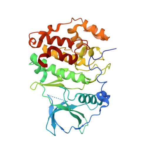
    The catalytic subunits of protein kinase CK2 are classified into two subtypes: CK2α1 and CK2α2. CK2α1 is an attractive drug-discovery target for various diseases such as cancers and nephritis. CK2α2 is defined as an off-target of CK2α1 and is a potential target in the development of male contraceptive drugs. High-resolution crystal structures of both isozymes are likely to provide crucial clues for the design of selective inhibitors of CK2α1 and/or CK2α2. To date, several crystal structures of CK2α1 have been solved at high resolutions of beyond 1.5 Å. However, crystal structures of CK2α2 have barely achieved a low resolution of around 3 Å because of the formation of needle-shaped crystals. In this study, new crystal forms were exploited and one provided a crystal structure of CK2α2 at 1.89 Å resolution. This result, together with the structure of CK2α1, will assist in the development of highly selective inhibitors for both isozymes.


  • Organizational Affiliation

    Graduate School of Science, Osaka Prefecture University, 1-1 Gakuen-cho, Naka-ku, Sakai, Osaka 599-8531, Japan.


Macromolecules
Find similar proteins by:  (by identity cutoff)  |  3D Structure
Entity ID: 1
MoleculeChains Sequence LengthOrganismDetailsImage
Casein kinase II subunit alpha'A [auth X],
B
339Homo sapiensMutation(s): 0 
Gene Names: CSNK2A2CK2A2
EC: 2.7.11.1
UniProt & NIH Common Fund Data Resources
Find proteins for P19784 (Homo sapiens)
Explore P19784 
Go to UniProtKB:  P19784
PHAROS:  P19784
GTEx:  ENSG00000070770 
Entity Groups  
Sequence Clusters30% Identity50% Identity70% Identity90% Identity95% Identity100% Identity
UniProt GroupP19784
Sequence Annotations
Expand
  • Reference Sequence
Small Molecules
Ligands 2 Unique
IDChains Name / Formula / InChI Key2D Diagram3D Interactions
NIO
Query on NIO

Download Ideal Coordinates CCD File 
C [auth X],
I [auth B]
NICOTINIC ACID
C6 H5 N O2
PVNIIMVLHYAWGP-UHFFFAOYSA-N
SO4
Query on SO4

Download Ideal Coordinates CCD File 
D [auth X]
E [auth X]
F [auth X]
G [auth X]
H [auth X]
D [auth X],
E [auth X],
F [auth X],
G [auth X],
H [auth X],
J [auth B],
K [auth B],
L [auth B]
SULFATE ION
O4 S
QAOWNCQODCNURD-UHFFFAOYSA-L
Experimental Data & Validation

Experimental Data

  • Method: X-RAY DIFFRACTION
  • Resolution: 1.89 Å
  • R-Value Free: 0.263 
  • R-Value Work: 0.211 
  • R-Value Observed: 0.214 
  • Space Group: P 1 21 1
Unit Cell:
Length ( Å )Angle ( ˚ )
a = 46.468α = 90
b = 102.422β = 95.02
c = 72.229γ = 90
Software Package:
Software NamePurpose
PHENIXrefinement
HKL-2000data reduction
HKL-2000data scaling
MOLREPphasing

Structure Validation

View Full Validation Report


Entry History 

Deposition Data

Revision History  (Full details and data files)

  • Version 1.0: 2018-05-16
    Type: Initial release
  • Version 1.1: 2023-11-22
    Changes: Data collection, Database references, Refinement description