A five-residue motif for the design of domain swapping in proteins.
Nandwani, N., Surana, P., Negi, H., Mascarenhas, N.M., Udgaonkar, J.B., Das, R., Gosavi, S.(2019) Nat Commun 10: 452-452
- PubMed: 30692525 Search on PubMedSearch on PubMed Central
- DOI: https://doi.org/10.1038/s41467-019-08295-x
- Primary Citation Related Structures:
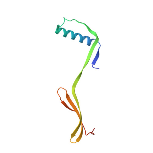
5YCT, 5YCU, 5YCW, 6IWJ - PubMed Abstract:
Domain swapping is the process by which identical monomeric proteins exchange structural elements to generate dimers/oligomers. Although engineered domain swapping is a compelling strategy for protein assembly, its application has been limited due to the lack of simple and reliable design approaches. Here, we demonstrate that the hydrophobic five-residue 'cystatin motif' (QVVAG) from the domain-swapping protein Stefin B, when engineered into a solvent-exposed, tight surface loop between two β-strands prevents the loop from folding back upon itself, and drives domain swapping in non-domain-swapping proteins. High-resolution structural studies demonstrate that engineering the QVVAG stretch independently into various surface loops of four structurally distinct non-domain-swapping proteins enabled the design of different modes of domain swapping in these proteins, including single, double and open-ended domain swapping. These results suggest that the introduction of the QVVAG motif can be used as a mutational approach for engineering domain swapping in diverse β-hairpin proteins.
- National Centre for Biological Sciences, Tata Institute of Fundamental Research, Bengaluru, 560065, India.
Organizational Affiliation: