5Y2J

Crystal structure of the oligomerization domain of NSP4 from rotavirus strain MF66

  • Classification: VIRAL PROTEIN
  • Organism(s): Bovine rotavirus G10
  • Expression System: Escherichia coli BL21(DE3)
  • Mutation(s): No 

  • Deposited: 2017-07-26 Released: 2018-03-14 
  • Deposition Author(s): Suguna, K., Kumar, S.
  • Funding Organization(s): Department of Biotechnology (DBT), Indian Institute of Science, (IISc) Partnership Program for Advanced Research in Biological Sciences and Bioengineering

Experimental Data Snapshot

  • Method: X-RAY DIFFRACTION
  • Resolution: 2.55 Å
  • R-Value Free: 0.258 
  • R-Value Work: 0.222 
  • R-Value Observed: 0.224 

wwPDB Validation   3D Report Full Report


Ligand Structure Quality Assessment 


This is version 1.2 of the entry. See complete history


Literature

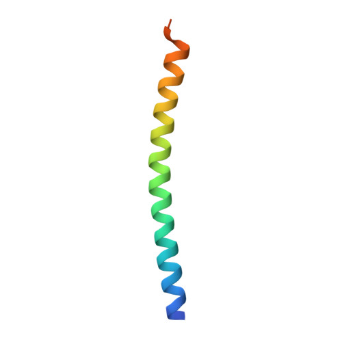
New tetrameric forms of the rotavirus NSP4 with antiparallel helices.

Kumar, S.Ramappa, R.Pamidimukkala, K.Rao, C.D.Suguna, K.

(2018) Arch Virol 163: 1531-1547

  • DOI: https://doi.org/10.1007/s00705-018-3753-6
  • Primary Citation of Related Structures:  
    5Y2E, 5Y2H, 5Y2J

  • PubMed Abstract: 

    Rotavirus nonstructural protein 4, the first viral enterotoxin to be identified, is a multidomain, multifunctional glycoprotein. Earlier, we reported a Ca 2+ -bound coiled-coil tetrameric structure of the diarrhea-inducing region of NSP4 from the rotavirus strains SA11 and I321 and a Ca 2+ -free pentameric structure from the rotavirus strain ST3, all with a parallel arrangement of α-helices. pH was found to determine the oligomeric state: a basic pH favoured a tetramer, whereas an acidic pH favoured a pentamer. Here, we report two novel forms of the coiled-coil region of NSP4 from the bovine rotavirus strains MF66 and NCDV. These crystallized at acidic pH, forming antiparallel coiled-coil tetrameric structures without any bound Ca 2+ ion. Structural and mutational studies of the coiled-coil regions of NSP4 revealed that the nature of the residue at position 131 (Tyr/His) plays an important role in the observed structural diversity.


  • Organizational Affiliation

    Molecular Biophysics Unit, Indian Institute of Science, Bangalore, India.


Macromolecules
Find similar proteins by:  (by identity cutoff)  |  3D Structure
Entity ID: 1
MoleculeChains Sequence LengthOrganismDetailsImage
Nonstructural protein 4
A, B, C, D
53Bovine rotavirus G10Mutation(s): 0 
UniProt
Find proteins for Q6QT01 (Bovine rotavirus G10)
Explore Q6QT01 
Go to UniProtKB:  Q6QT01
Entity Groups  
Sequence Clusters30% Identity50% Identity70% Identity90% Identity95% Identity100% Identity
UniProt GroupQ6QT01
Sequence Annotations
Expand
  • Reference Sequence
Experimental Data & Validation

Experimental Data

  • Method: X-RAY DIFFRACTION
  • Resolution: 2.55 Å
  • R-Value Free: 0.258 
  • R-Value Work: 0.222 
  • R-Value Observed: 0.224 
  • Space Group: P 21 21 21
Unit Cell:
Length ( Å )Angle ( ˚ )
a = 53.61α = 90
b = 69.57β = 90
c = 69.57γ = 90
Software Package:
Software NamePurpose
PHENIXrefinement
iMOSFLMdata processing
Aimlessdata scaling
CRANK2phasing

Structure Validation

View Full Validation Report


Ligand Structure Quality Assessment 


Entry History & Funding Information

Deposition Data


Funding OrganizationLocationGrant Number
Department of Biotechnology (DBT), Indian Institute of Science, (IISc) Partnership Program for Advanced Research in Biological Sciences and BioengineeringIndia--

Revision History  (Full details and data files)

  • Version 1.0: 2018-03-14
    Type: Initial release
  • Version 1.1: 2018-05-30
    Changes: Data collection, Database references
  • Version 1.2: 2024-03-27
    Changes: Data collection, Database references, Derived calculations