5XLI

Structure of anti-Angiotensin II type2 receptor antibody (D5711-4A03)


Experimental Data Snapshot

  • Method: X-RAY DIFFRACTION
  • Resolution: 1.70 Å
  • R-Value Free: 0.216 
  • R-Value Work: 0.179 
  • R-Value Observed: 0.181 

wwPDB Validation   3D Report Full Report


This is version 1.2 of the entry. See complete history


Literature

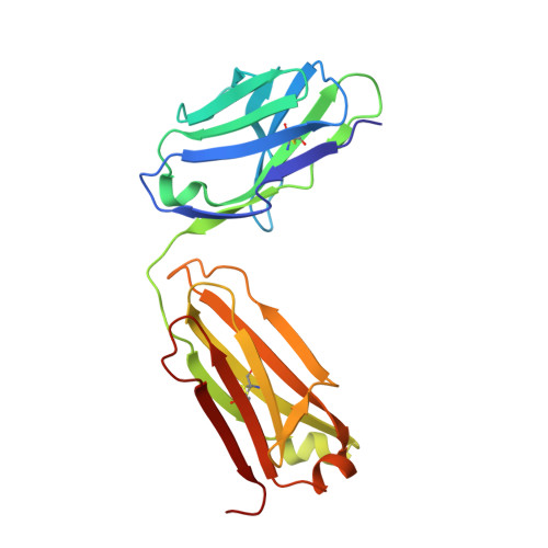
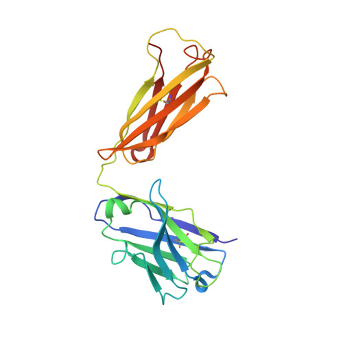
Crystal structure of the human angiotensin II type 2 receptor bound to an angiotensin II analog.

Asada, H.Horita, S.Hirata, K.Shiroishi, M.Shiimura, Y.Iwanari, H.Hamakubo, T.Shimamura, T.Nomura, N.Kusano-Arai, O.Uemura, T.Suno, C.Kobayashi, T.Iwata, S.

(2018) Nat Struct Mol Biol 25: 570-576

  • DOI: https://doi.org/10.1038/s41594-018-0079-8
  • Primary Citation of Related Structures:  
    5XJM, 5XLI

  • PubMed Abstract: 

    Angiotensin II (AngII) plays a central role in regulating human blood pressure, which is mainly mediated by interactions between AngII and the G-protein-coupled receptors (GPCRs) AngII type 1 receptor (AT 1 R) and AngII type 2 receptor (AT 2 R). We have solved the crystal structure of human AT 2 R binding the peptide ligand [Sar 1 , Ile 8 ]AngII and its specific antibody at 3.2-Å resolution. [Sar 1 , Ile 8 ]AngII interacts with both the 'core' binding domain, where the small-molecule ligands of AT 1 R and AT 2 R bind, and the 'extended' binding domain, which is equivalent to the allosteric modulator binding site of muscarinic acetylcholine receptor. We generated an antibody fragment to stabilize the extended binding domain that functions as a positive allosteric modulator. We also identified a signature positively charged cluster, which is conserved among peptide-binding receptors, to locate C termini at the bottom of the binding pocket. The reported results should help with designing ligands for angiotensin receptors and possibly to other peptide GPCRs.


  • Organizational Affiliation

    Department of Cell Biology, Graduate School of Medicine, Kyoto University, Kyoto, Japan.


Macromolecules
Find similar proteins by:  (by identity cutoff)  |  3D Structure
Entity ID: 1
MoleculeChains Sequence LengthOrganismDetailsImage
FabLA [auth a],
B [auth b]
213Mus musculusMutation(s): 0 
Entity Groups  
Sequence Clusters30% Identity50% Identity70% Identity90% Identity95% Identity100% Identity
Sequence Annotations
Expand
  • Reference Sequence
Find similar proteins by:  (by identity cutoff)  |  3D Structure
Entity ID: 2
MoleculeChains Sequence LengthOrganismDetailsImage
FabHC [auth c],
D [auth d]
220Mus musculusMutation(s): 0 
Entity Groups  
Sequence Clusters30% Identity50% Identity70% Identity90% Identity95% Identity100% Identity
Sequence Annotations
Expand
  • Reference Sequence
Experimental Data & Validation

Experimental Data

  • Method: X-RAY DIFFRACTION
  • Resolution: 1.70 Å
  • R-Value Free: 0.216 
  • R-Value Work: 0.179 
  • R-Value Observed: 0.181 
  • Space Group: P 1 21 1
Unit Cell:
Length ( Å )Angle ( ˚ )
a = 41.654α = 90
b = 150.498β = 93.1
c = 75.322γ = 90
Software Package:
Software NamePurpose
PHENIXrefinement
XSCALEdata scaling
PDB_EXTRACTdata extraction

Structure Validation

View Full Validation Report


Entry History & Funding Information

Deposition Data


Funding OrganizationLocationGrant Number
Japan Agency for Medical Research and DevelopmentJapanA19170600003
Japan Society for the Promotion of ScienceJapan15J04343
Japan Society for the Promotion of ScienceJapan22590270

Revision History  (Full details and data files)

  • Version 1.0: 2018-07-11
    Type: Initial release
  • Version 1.1: 2018-07-25
    Changes: Data collection, Database references
  • Version 1.2: 2023-11-22
    Changes: Data collection, Database references, Refinement description