Crystal structure and DNA-binding property of the ATPase domain of bacterial mismatch repair endonuclease MutL from Aquifex aeolicus
Fukui, K., Iino, H., Baba, S., Kumasaka, T., Kuramitsu, S., Yano, T.(2017) Biochim Biophys Acta 1865: 1178-1187
- PubMed: 28668638 Search on PubMed
- DOI: https://doi.org/10.1016/j.bbapap.2017.06.024
- Primary Citation Related Structures:
5X9Y - PubMed Abstract:
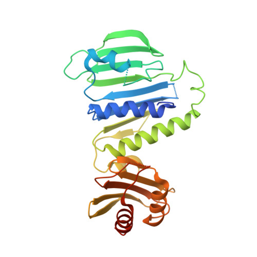
DNA mismatch repair (MMR) system corrects mismatched bases that are generated mainly by DNA replication errors. The repair system excises the error-containing single-stranded region and enables the re-synthesis of the strand. In the early reactions of MMR, MutL endonuclease incises the newly-synthesized/error-containing strand of the duplex to initiate the downstream excision reaction. MutL endonuclease consists of the N-terminal ATPase and C-terminal endonuclease domains. In this study, we report the crystal structure of the ATPase domain of MutL endonuclease from Aquifex aeolicus. The overall structure of the domain was similar to those of human MutL homologs and Escherichia coli MutL, although E. coli MutL has no endonuclease activity. The ATPase domain was comprised of two subdomains: the N-terminal ATP-binding subdomain and the C-terminal α-β sandwich subdomain. Site-directed mutagenesis experiment identified DNA-interacting eight basic amino acid residues, which were distributed across both the two subdomains and formed a DNA-binding cleft. Docking simulation between the structures of the ATPase and endonuclease domains generated a reliable model structure for the full-length A. aeolicus MutL, which satisfies our previous result of small-angle X-ray scattering analysis. On the basis of the model structure and further experimental results, we concluded that the two separate DNA-binding sites in the full-length A. aeolicus MutL simultaneously bind a dsDNA molecule.
- Department of Biochemistry, Osaka Medical College, 2-7 Daigakumachi, Takatsuki, Osaka 569-8686, Japan. Electronic address: k.fukui@osaka-med.ac.jp.
Organizational Affiliation: