5X93

Human endothelin receptor type-B in complex with antagonist K-8794


Experimental Data Snapshot

  • Method: X-RAY DIFFRACTION
  • Resolution: 2.20 Å
  • R-Value Free: 0.239 
  • R-Value Work: 0.211 
  • R-Value Observed: 0.212 

wwPDB Validation   3D Report Full Report


Ligand Structure Quality Assessment 


This is version 1.2 of the entry. See complete history


Literature

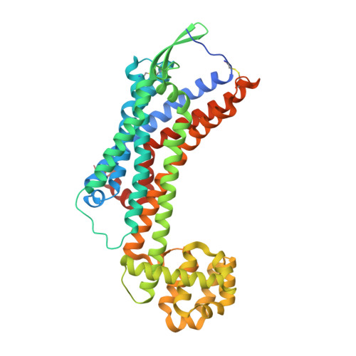
X-ray structures of endothelin ETB receptor bound to clinical antagonist bosentan and its analog

Shihoya, W.Nishizawa, T.Yamashita, K.Inoue, A.Hirata, K.Kadji, F.M.N.Okuta, A.Tani, K.Aoki, J.Fujiyoshi, Y.Doi, T.Nureki, O.

(2017) Nat Struct Mol Biol 24: 758-764

  • DOI: https://doi.org/10.1038/nsmb.3450
  • Primary Citation of Related Structures:  
    5X93, 5XPR

  • PubMed Abstract: 
  • Endothelin receptors (ETRs) have crucial roles in vascular control and are targets for drugs designed to treat circulatory-system diseases and cancer progression. The nonpeptide dual-ETR antagonist bosentan is the first oral drug approved to treat pulmonary arterial hypertension ...

    Endothelin receptors (ETRs) have crucial roles in vascular control and are targets for drugs designed to treat circulatory-system diseases and cancer progression. The nonpeptide dual-ETR antagonist bosentan is the first oral drug approved to treat pulmonary arterial hypertension. Here we report crystal structures of human endothelin ET B receptor bound to bosentan and to the ET B -selective analog K-8794, at 3.6-Å and 2.2-Å resolution, respectively. The K-8794-bound structure reveals the detailed water-mediated hydrogen-bonding network at the transmembrane core, which could account for the weak negative allosteric modulation of ET B by Na + ions. The bosentan-bound structure reveals detailed interactions with ET B , which are probably conserved in the ET A receptor. A comparison of the two structures shows unexpected similarity between antagonist and agonist binding. Despite this similarity, bosentan sterically prevents the inward movement of transmembrane helix 6 (TM6), and thus exerts its antagonistic activity. These structural insights will facilitate the rational design of new ETR-targeting drugs.


    Organizational Affiliation

    Department of Biophysics and Biochemistry, Graduate School of Science, The University of Tokyo, Tokyo, Japan.


Macromolecules
Find similar proteins by:  (by identity cutoff)  |  3D Structure
Entity ID: 1
MoleculeChainsSequence LengthOrganismDetailsImage
Endothelin B receptor,Endolysin,Endothelin B receptorA464Homo sapiensTequatrovirus T4Mutation(s): 10 
Gene Names: EDNRBETRBE
EC: 3.2.1.17
Membrane Entity: Yes 
UniProt & NIH Common Fund Data Resources
Find proteins for P00720 (Enterobacteria phage T4)
Explore P00720 
Go to UniProtKB:  P00720
Find proteins for P24530 (Homo sapiens)
Explore P24530 
Go to UniProtKB:  P24530
PHAROS:  P24530
Entity Groups  
Sequence Clusters30% Identity50% Identity70% Identity90% Identity95% Identity100% Identity
UniProt GroupsP00720P24530
Protein Feature View
Expand
  • Reference Sequence
Small Molecules
Ligands 4 Unique
IDChainsName / Formula / InChI Key2D Diagram3D Interactions
K87
Query on K87

Download Ideal Coordinates CCD File 
B [auth A]3-[6-[(4-tert-butylphenyl)sulfonylamino]-5-(2-methoxyphenoxy)-2-pyrimidin-2-yl-pyrimidin-4-yl]oxy-N-(2,6-dimethylphenyl)propanamide
C36 H38 N6 O6 S
MTNFANNLCUMCNB-UHFFFAOYSA-N
 Ligand Interaction
CLR
Query on CLR

Download Ideal Coordinates CCD File 
N [auth A]CHOLESTEROL
C27 H46 O
HVYWMOMLDIMFJA-DPAQBDIFSA-N
 Ligand Interaction
OLC
Query on OLC

Download Ideal Coordinates CCD File 
C [auth A],
D [auth A],
E [auth A],
F [auth A],
G [auth A],
C [auth A],
D [auth A],
E [auth A],
F [auth A],
G [auth A],
H [auth A],
I [auth A],
J [auth A],
K [auth A],
L [auth A],
M [auth A]
(2R)-2,3-dihydroxypropyl (9Z)-octadec-9-enoate
C21 H40 O4
RZRNAYUHWVFMIP-GDCKJWNLSA-N
 Ligand Interaction
SO4
Query on SO4

Download Ideal Coordinates CCD File 
O [auth A],
P [auth A],
Q [auth A],
R [auth A],
S [auth A]
SULFATE ION
O4 S
QAOWNCQODCNURD-UHFFFAOYSA-L
 Ligand Interaction
Binding Affinity Annotations 
IDSourceBinding Affinity
K87 Binding MOAD:  5X93 IC50: 150 (nM) from 1 assay(s)
Experimental Data & Validation

Experimental Data

  • Method: X-RAY DIFFRACTION
  • Resolution: 2.20 Å
  • R-Value Free: 0.239 
  • R-Value Work: 0.211 
  • R-Value Observed: 0.212 
  • Space Group: C 2 2 21
Unit Cell:
Length ( Å )Angle ( ˚ )
a = 74.13α = 90
b = 147.48β = 90
c = 108.38γ = 90
Software Package:
Software NamePurpose
PHENIXrefinement
XSCALEdata scaling
PDB_EXTRACTdata extraction
XDSdata reduction
PHASERphasing

Structure Validation

View Full Validation Report


Ligand Structure Quality Assessment 


Entry History 

Deposition Data

Revision History  (Full details and data files)

  • Version 1.0: 2017-08-16
    Type: Initial release
  • Version 1.1: 2017-09-20
    Changes: Database references
  • Version 1.2: 2017-11-15
    Changes: Structure summary