5X4U

PAC from Oscillatoriaacuminata after 60 seconds photoactivation


Experimental Data Snapshot

  • Method: X-RAY DIFFRACTION
  • Resolution: 1.80 Å
  • R-Value Free: 0.245 
  • R-Value Work: 0.203 
  • R-Value Observed: 0.205 

wwPDB Validation   3D Report Full Report


Ligand Structure Quality Assessment 


This is version 1.2 of the entry. See complete history


Literature

Molecular mechanism of photoactivation of a light-regulated adenylate cyclase.

Ohki, M.Sato-Tomita, A.Matsunaga, S.Iseki, M.Tame, J.R.H.Shibayama, N.Park, S.Y.

(2017) Proc Natl Acad Sci U S A 114: 8562-8567

  • DOI: https://doi.org/10.1073/pnas.1704391114
  • Primary Citation of Related Structures:  
    5X4T, 5X4U, 5X4V

  • PubMed Abstract: 

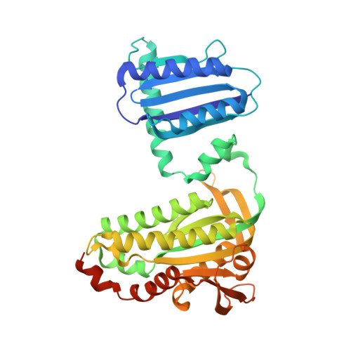
    The photoactivated adenylate cyclase (PAC) from the photosynthetic cyanobacterium Oscillatoria acuminata (OaPAC) detects light through a flavin chromophore within the N-terminal BLUF domain. BLUF domains have been found in a number of different light-activated proteins, but with different relative orientations. The two BLUF domains of OaPAC are found in close contact with each other, forming a coiled coil at their interface. Crystallization does not impede the activity switching of the enzyme, but flash cooling the crystals to cryogenic temperatures prevents the signature spectral changes that occur on photoactivation/deactivation. High-resolution crystallographic analysis of OaPAC in the fully activated state has been achieved by cryocooling the crystals immediately after light exposure. Comparison of the isomorphous light- and dark-state structures shows that the active site undergoes minimal changes, yet enzyme activity may increase up to 50-fold, depending on conditions. The OaPAC models will assist the development of simple, direct means to raise the cyclic AMP levels of living cells by light, and other tools for optogenetics.


  • Organizational Affiliation

    Drug Design Laboratory, Graduate School of Medical Life Science, Yokohama City University, Tsurumi, Yokohama 230-0045, Japan.


Macromolecules
Find similar proteins by:  (by identity cutoff)  |  3D Structure
Entity ID: 1
MoleculeChains Sequence LengthOrganismDetailsImage
Photoactivated adenylyl cyclase366CyanobacteriotaMutation(s): 0 
UniProt
Find proteins for K9TLZ5 (Oscillatoria acuminata PCC 6304)
Explore K9TLZ5 
Go to UniProtKB:  K9TLZ5
Entity Groups  
Sequence Clusters30% Identity50% Identity70% Identity90% Identity95% Identity100% Identity
UniProt GroupK9TLZ5
Sequence Annotations
Expand
  • Reference Sequence
Small Molecules
Ligands 1 Unique
IDChains Name / Formula / InChI Key2D Diagram3D Interactions
FMN
Query on FMN

Download Ideal Coordinates CCD File 
B [auth A]FLAVIN MONONUCLEOTIDE
C17 H21 N4 O9 P
FVTCRASFADXXNN-SCRDCRAPSA-N
Experimental Data & Validation

Experimental Data

  • Method: X-RAY DIFFRACTION
  • Resolution: 1.80 Å
  • R-Value Free: 0.245 
  • R-Value Work: 0.203 
  • R-Value Observed: 0.205 
  • Space Group: P 61 2 2
Unit Cell:
Length ( Å )Angle ( ˚ )
a = 77.149α = 90
b = 77.149β = 90
c = 204.824γ = 120
Software Package:
Software NamePurpose
PHENIXrefinement
HKL-2000data reduction
HKL-2000data scaling
MOLREPmodel building

Structure Validation

View Full Validation Report


Ligand Structure Quality Assessment 


Entry History 

Deposition Data

Revision History  (Full details and data files)

  • Version 1.0: 2017-07-26
    Type: Initial release
  • Version 1.1: 2017-12-06
    Changes: Database references
  • Version 1.2: 2024-03-27
    Changes: Data collection, Database references