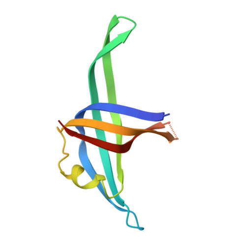
Insight into the interaction between PriB and DnaT on bacterial DNA replication restart: Significance of the residues on PriB dimer interface and highly acidic region on DnaT.
Fujiyama, S., Abe, Y., Shiroishi, M., Ikeda, Y., Ueda, T.(2019) Biochim Biophys Acta Proteins Proteom 1867: 367-375
- PubMed: 30659961
- DOI: https://doi.org/10.1016/j.bbapap.2019.01.008
- Primary Citation of Related Structures:
5WQV - PubMed Abstract:
When the replisome collapses at a DNA damage site, a sequence-independent replication restart system is required. In Escherichia coli, PriA, PriB, and DnaT assemble in an orderly fashion at the stalled replication fork and achieve the reloading of the replisome. PriB-DnaT interaction is considered a significant step in the replication restart. In this study, we examined the contribution of the residues Ser20, His26 and Ser55, which are located on the PriB dimer interface. These residues are proximal to Glu39 and Arg44, which are important for PriB-DnaT interaction. Mutational analyses revealed that His26 and Ser20 of PriB are important for the interaction with DnaT, and that the Ser55 residue of PriB might have a role in negatively regulating the DnaT binding. These residues are involved in not only the interaction between PriB and DnaT but also the dissociation of single-stranded DNA (ssDNA) from the PriB-ssDNA complex due to DnaT binding. Moreover, NMR study indicates that the region Asp66-Glu76 on the linker between DnaT domains is involved in the interaction with wild-type PriB. These findings provide significant information about the molecular mechanism underlying replication restart in bacteria.
- Laboratory of Protein Structure, Function and Design, Graduate School of Pharmaceutical Sciences, Kyushu University, 3-1-1 Maidashi, Higashi-ku, Fukuoka 812-8582, Japan; Research Fellow of Japan Society for the Promotion of Science, 5-3-1 Koujimachi, Chiyoda-ku, Tokyo 102-0083, Japan.
Organizational Affiliation:


