Crystal Structure of Cocosin, A Potential Food Allergen from Coconut (Cocos nucifera)
Jin, T., Wang, C., Zhang, C., Wang, Y., Chen, Y.W., Guo, F., Howard, A., Cao, M.J., Fu, T.J., McHugh, T.H., Zhang, Y.(2017) J Agric Food Chem 65: 7560-7568
- PubMed: 28712292 Search on PubMed
- DOI: https://doi.org/10.1021/acs.jafc.7b02252
- Primary Citation Related Structures:
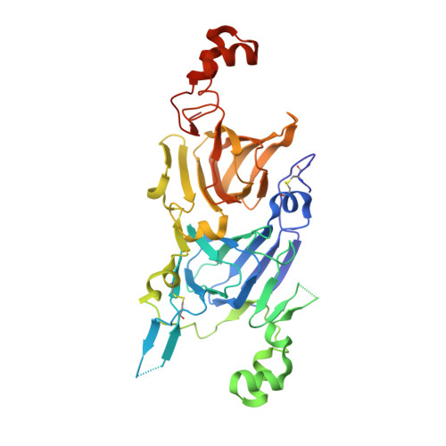
5WPW - PubMed Abstract:
Coconut (Cocos nucifera) is an important palm tree. Coconut fruit is widely consumed. The most abundant storage protein in coconut fruit is cocosin (a likely food allergen), which belongs to the 11S globulin family. Cocosin was crystallized near a century ago, but its structure remains unknown. By optimizing crystallization conditions and cryoprotectant solutions, we were able to obtain cocosin crystals that diffracted to 1.85 Å. The cocosin gene was cloned from genomic DNA isolated from dry coconut tissue. The protein sequence deduced from the predicted cocosin coding sequence was used to guide model building and structure refinement. The structure of cocosin was determined for the first time, and it revealed a typical 11S globulin feature of a double layer doughnut-shaped hexamer.
- Laboratory of Structural Immunology, CAS Key Laboratory of Innate Immunity and Chronic Diseases, CAS Center for Excellence in Molecular Cell Sciences, School of Life Sciences and Medical Center, University of Science and Technology of China , Hefei 230027 China.
Organizational Affiliation: