Identification of a Unique Inhibitor-Binding Site on Choline Kinase alpha.
Kall, S.L., Delikatny, E.J., Lavie, A.(2018) Biochemistry 57: 1316-1325
- PubMed: 29389115 Search on PubMed
- DOI: https://doi.org/10.1021/acs.biochem.7b01257
- Primary Citation Related Structures:
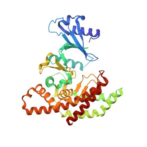
5W6O - PubMed Abstract:
Choline kinase α (ChoKα) is an enzyme that is upregulated in many types of cancer and has been shown to be tumorigenic. As such, it makes a promising target for inhibiting tumor growth. Though there have been several inhibitors synthesized for ChoKα, not all of them demonstrate the same efficacy in vivo, though the reasons behind this difference in potency are not clear. One particular inhibitor, designated TCD-717, has recently completed phase I clinical trials. Cell culture and in vitro studies support the powerful inhibitory effect TCD-717 has on ChoKα, but an examination of the inhibitor's interaction with the ChoKα enzyme has been missing prior to this work. Here we detail the 2.35 Å structure of ChoKα in complex with TCD-717. Examination of this structure in conjunction with kinetic assays reveals that TCD-717 does not bind directly in the choline pocket as do previously characterized ChoKα inhibitors, but rather in a proximal but novel location near the surface of the enzyme. The unique binding site identified for TCD-717 lends insight for the future design of more potent in vivo inhibitors for ChoKα.
- Department of Biochemistry and Molecular Genetics, University of Illinois at Chicago , Chicago, Illinois 60607, United States.
Organizational Affiliation: