5VYP | pdb_00005vyp

Crystal structure of the Plant Defensin NsD7 bound to PIP2


Experimental Data Snapshot

  • Method: X-RAY DIFFRACTION
  • Resolution: 2.60 Å
  • R-Value Free: 
    0.270 (Depositor), 0.269 (DCC) 
  • R-Value Work: 
    0.214 (Depositor), 0.216 (DCC) 
  • R-Value Observed: 
    0.217 (Depositor) 

Starting Model: experimental
View more details

wwPDB Validation 3D Report Full Report

Validation slider image for 5VYP

Ligand Structure Quality Assessment 


This is version 1.5 of the entry. See complete history

Literature

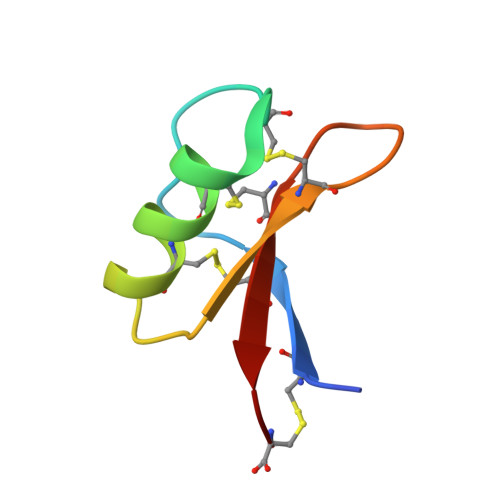
Structure of the defensin NsD7 in complex with PIP2 reveals that defensin : lipid oligomer topologies are dependent on lipid type.

Jarva, M.Lay, F.T.Hulett, M.D.Kvansakul, M.

(2017) FEBS Lett 591: 2482-2490

  • DOI: https://doi.org/10.1002/1873-3468.12761
  • Primary Citation Related Structures: 
    5VYP

  • PubMed Abstract: 

    Defensins are innate immune molecules that upon recognition of specific phospholipids can disrupt microbial membranes by forming oligomeric assemblies. Structures of two related plant defensins, NaD1 and NsD7, bound to phosphatidylinositol 4,5-bisphosphate (PIP 2 ) and phosphatidic acid (PA), respectively, revealed striking differences in their oligomeric topologies. To understand how NsD7 binds different phospholipids and rationalize the different topologies, we determined the structure of an NsD7-PIP 2 complex. This structure reveals fundamental differences in phospholipid binding compared to NsD7-PA, and an oligomeric topology nearly identical to the previously determined NaD1-PIP 2 complex, establishing that the PIP 2 fibril topology is conserved between NaD1 and NsD7. Our findings highlight the remarkable ability of defensins to bind different types of phospholipids to form oligomeric fibrils with diverse topologies.


  • Organizational Affiliation
    • Department of Biochemistry and Genetics, La Trobe Institute for Molecular Science, La Trobe University, Melbourne, Vic, Australia.

Macromolecule Content 

  • Total Structure Weight: 151.09 kDa 
  • Atom Count: 10,341 
  • Modeled Residue Count: 1,145 
  • Deposited Residue Count: 1,152 
  • Unique protein chains: 1

Macromolecules

Find similar proteins by:|  3D Structure
Entity ID: 1
MoleculeChains  Sequence LengthOrganismDetailsImage
Defensin NsD7
A, B, C, D, E
A, B, C, D, E, F, G, H, I, J, K, L, M, N, O, P, Q, R, S, T, U, V, W, X
48Nicotiana suaveolensMutation(s): 0 
UniProt
Find proteins for C0HK49 (Nicotiana suaveolens)
Explore C0HK49 
Go to UniProtKB:  C0HK49
Entity Groups
Sequence Clusters30% Identity50% Identity70% Identity90% Identity95% Identity100% Identity
UniProt GroupC0HK49
Sequence Annotations
Expand
Reference Sequence

Small Molecules

Ligands 3 Unique
IDChains Name / Formula / InChI Key2D Diagram3D Interactions
PIO

Query on PIO


Download:Ideal Coordinates CCD File
BA [auth B]
BB [auth S]
CA [auth C]
DB [auth T]
EA [auth D]
BA [auth B],
BB [auth S],
CA [auth C],
DB [auth T],
EA [auth D],
FB [auth U],
GA [auth E],
HA [auth F],
IA [auth G],
KA [auth H],
LA [auth I],
LB [auth V],
MB [auth W],
NA [auth J],
OA [auth K],
PA [auth L],
PB [auth X],
QA [auth M],
TA [auth N],
UA [auth O],
VA [auth P],
XA [auth Q],
Y [auth A],
ZA [auth R]
[(2R)-2-octanoyloxy-3-[oxidanyl-[(1R,2R,3S,4R,5R,6S)-2,3,6-tris(oxidanyl)-4,5-diphosphonooxy-cyclohexyl]oxy-phosphoryl]oxy-propyl] octanoate
C25 H49 O19 P3
XLNCEHRXXWQMPK-MJUMVPIBSA-N
SO4

Query on SO4


Download:Ideal Coordinates CCD File
AA [auth A]
AB [auth R]
FA [auth D]
GB [auth U]
HB [auth U]
AA [auth A],
AB [auth R],
FA [auth D],
GB [auth U],
HB [auth U],
IB [auth U],
JA [auth G],
Z [auth A]
SULFATE ION
O4 S
QAOWNCQODCNURD-UHFFFAOYSA-L
GOL

Query on GOL


Download:Ideal Coordinates CCD File
CB [auth S]
DA [auth C]
EB [auth T]
JB [auth U]
KB [auth U]
CB [auth S],
DA [auth C],
EB [auth T],
JB [auth U],
KB [auth U],
MA [auth I],
NB [auth W],
OB [auth W],
RA [auth M],
SA [auth M],
WA [auth P],
YA [auth Q]
GLYCEROL
C3 H8 O3
PEDCQBHIVMGVHV-UHFFFAOYSA-N

Experimental Data & Validation

Experimental Data

  • Method: X-RAY DIFFRACTION
  • Resolution: 2.60 Å
  • R-Value Free:  0.270 (Depositor), 0.269 (DCC) 
  • R-Value Work:  0.214 (Depositor), 0.216 (DCC) 
  • R-Value Observed: 0.217 (Depositor) 
Space Group: P 1 21 1
Unit Cell:
Length ( Å )Angle ( ˚ )
a = 75.605α = 90
b = 83.489β = 93.99
c = 105.194γ = 90
Software Package:
Software NamePurpose
PHENIXrefinement
Aimlessdata scaling
PHASERphasing
PDB_EXTRACTdata extraction
XDSdata reduction

Structure Validation

View Full Validation Report


Ligand Structure Quality Assessment 


Entry History 

& Funding Information

Deposition Data


Funding OrganizationLocationGrant Number
Australian Research Council (ARC)AustraliaFT130101349

Revision History  (Full details and data files)

  • Version 1.0: 2017-08-02
    Type: Initial release
  • Version 1.1: 2017-09-13
    Changes: Database references
  • Version 1.2: 2017-09-27
    Changes: Author supporting evidence
  • Version 1.3: 2020-01-01
    Changes: Author supporting evidence
  • Version 1.4: 2023-10-04
    Changes: Data collection, Database references, Refinement description
  • Version 1.5: 2024-11-20
    Changes: Structure summary