Purification and Characterization of RhoPDE, a Retinylidene/Phosphodiesterase Fusion Protein and Potential Optogenetic Tool from the Choanoflagellate Salpingoeca rosetta.
Lamarche, L.B., Kumar, R.P., Trieu, M.M., Devine, E.L., Cohen-Abeles, L.E., Theobald, D.L., Oprian, D.D.(2017) Biochemistry 56: 5812-5822
- PubMed: 28976747 Search on PubMedSearch on PubMed Central
- DOI: https://doi.org/10.1021/acs.biochem.7b00519
- Primary Citation Related Structures:
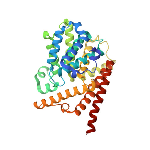
5VYD - PubMed Abstract:
RhoPDE is a type I rhodopsin/phosphodiesterase gene fusion product from the choanoflagellate Salpingoeca rosetta. The gene was discovered around the time that a similar type I rhodopsin/guanylyl cyclase fusion protein, RhoGC, was shown to control phototaxis of an aquatic fungus through a cGMP signaling pathway. RhoPDE has potential as an optogenetic tool catalyzing the hydrolysis of cyclic nucleotides. Here we provide an expression and purification system for RhoPDE, as well as a crystal structure of the C-terminal phosphodiesterase catalytic domain. We show that RhoPDE contains an even number of transmembrane segments, with N- and C-termini both located on the cytoplasmic surface of the cell membrane. The purified protein exhibits an absorption maximum at 490 nm in the dark state, which shifts to 380 nm upon exposure to light. The protein acts as a cGMP-selective phosphodiesterase. However, the activity does not appear to be modulated by light. The protein is also active with cAMP as a substrate, but with a roughly 5-7-fold lower k cat . A truncation consisting solely of the phosphodiesterase domain is also active with a k cat for cGMP roughly 6-9-fold lower than that of the full-length protein. The isolated PDE domain was crystallized, and the X-ray structure showed the protein to be a dimer similar to human PDE9. We anticipate that the purification system introduced here will enable further structural and biochemical experiments to improve our understanding of the function and mechanism of this unique fusion protein.
- Department of Biochemistry, Brandeis University , Waltham, Massachusetts 02454, United States.
Organizational Affiliation: