5VX8

VP8* of P[6] Human Rotavirus RV3


Experimental Data Snapshot

  • Method: X-RAY DIFFRACTION
  • Resolution: 2.00 Å
  • R-Value Free: 0.258 
  • R-Value Work: 0.208 
  • R-Value Observed: 0.211 

wwPDB Validation   3D Report Full Report


This is version 1.2 of the entry. See complete history


Literature

Glycan recognition in globally dominant human rotaviruses.

Hu, L.Sankaran, B.Laucirica, D.R.Patil, K.Salmen, W.Ferreon, A.C.M.Tsoi, P.S.Lasanajak, Y.Smith, D.F.Ramani, S.Atmar, R.L.Estes, M.K.Ferreon, J.C.Prasad, B.V.V.

(2018) Nat Commun 9: 2631-2631

  • DOI: https://doi.org/10.1038/s41467-018-05098-4

  • PubMed Abstract: 

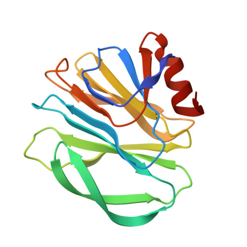
    Rotaviruses (RVs) cause life-threatening diarrhea in infants and children worldwide. Recent biochemical and epidemiological studies underscore the importance of histo-blood group antigens (HBGA) as both cell attachment and susceptibility factors for the globally dominant P[4], P[6], and P[8] genotypes of human RVs. How these genotypes interact with HBGA is not known. Here, our crystal structures of P[4] and a neonate-specific P[6] VP8*s alone and in complex with H-type I HBGA reveal a unique glycan binding site that is conserved in the globally dominant genotypes and allows for the binding of ABH HBGAs, consistent with their prevalence. Remarkably, the VP8* of P[6] RVs isolated from neonates displays subtle structural changes in this binding site that may restrict its ability to bind branched glycans. This provides a structural basis for the age-restricted tropism of some P[6] RVs as developmentally regulated unbranched glycans are more abundant in the neonatal gut.


  • Organizational Affiliation

    Verna and Marrs McLean Department of Biochemistry and Molecular Biology, Baylor College of Medicine, Houston, TX, 77030, USA.


Macromolecules
Find similar proteins by:  (by identity cutoff)  |  3D Structure
Entity ID: 1
MoleculeChains Sequence LengthOrganismDetailsImage
Outer capsid protein VP4
A, B
161Human rotavirus AMutation(s): 0 
UniProt
Find proteins for D7F7M7 (Human rotavirus A)
Explore D7F7M7 
Go to UniProtKB:  D7F7M7
Entity Groups  
Sequence Clusters30% Identity50% Identity70% Identity90% Identity95% Identity100% Identity
UniProt GroupD7F7M7
Sequence Annotations
Expand
  • Reference Sequence
Experimental Data & Validation

Experimental Data

  • Method: X-RAY DIFFRACTION
  • Resolution: 2.00 Å
  • R-Value Free: 0.258 
  • R-Value Work: 0.208 
  • R-Value Observed: 0.211 
  • Space Group: P 1
Unit Cell:
Length ( Å )Angle ( ˚ )
a = 27.79α = 94.86
b = 43.1β = 99.4
c = 65.74γ = 90.22
Software Package:
Software NamePurpose
PHENIXrefinement
iMOSFLMdata reduction
SCALAdata scaling
PHASERphasing

Structure Validation

View Full Validation Report


Entry History & Funding Information

Deposition Data


Funding OrganizationLocationGrant Number
National Institutes of Health/National Institute Of Allergy and Infectious Diseases (NIH/NIAID)United StatesAI36040
Robert A. Welch FoundationUnited StatesQ1279

Revision History  (Full details and data files)

  • Version 1.0: 2018-07-18
    Type: Initial release
  • Version 1.1: 2019-12-11
    Changes: Author supporting evidence
  • Version 1.2: 2023-10-04
    Changes: Data collection, Database references, Refinement description