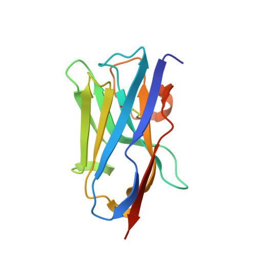
Structure and specificity of several triclocarban-binding single domain camelid antibody fragments.
Tabares-da Rosa, S., Wogulis, L.A., Wogulis, M.D., Gonzalez-Sapienza, G., Wilson, D.K.(2019) J Mol Recognit 32: e2755-e2755
- PubMed: 30033524 Search on PubMedSearch on PubMed Central
- DOI: https://doi.org/10.1002/jmr.2755
- Primary Citation Related Structures:
5VL2, 5VLV, 5VM0, 5VM4, 5VM6 - PubMed Abstract:
The variable VHH domains of camelid single chain antibodies have been useful in numerous biotechnology applications due to their simplicity, biophysical properties, and abilities to bind to their cognate antigens with high affinities and specificity. Their interactions with proteins have been well-studied, but considerably less work has been done to characterize their ability to bind haptens. A high-resolution structural study of three nanobodies (T4, T9, and T10) which have been shown to bind triclocarban (TCC, 3-(4-chlorophenyl)-1-(3,4-dichlorophenyl)urea) with near-nanomolar affinity shows that binding occurs in a tunnel largely formed by CDR1 rather than a surface or lateral binding mode seen in other nanobody-hapten interactions. Additional significant interactions are formed with a non-hypervariable loop, sometimes dubbed "CDR4". A comparison of apo and holo forms of T9 and T10 shows that the binding site undergoes little conformational change upon binding of TCC. Structures of three nanobody-TCC complexes demonstrated there was not a standard binding mode. T4 and T9 have a high degree of sequence identity and bind the hapten in a nearly identical manner, while the more divergent T10 binds TCC in a slightly displaced orientation with the urea moiety rotated approximately 180° along the long axis of the molecule. In addition to methotrexate, this is the second report of haptens binding in a tunnel formed by CDR1, suggesting that compounds with similar hydrophobicity and shape could be recognized by nanobodies in analogous fashion. Structure-guided mutations failed to improve binding affinity for T4 and T9 underscoring the high degree of natural optimization.
- Cátedra de Inmunología, Facultad de Química, Instituto de Higiene, UDELAR, Uruguay.
Organizational Affiliation: