5VHV | pdb_00005vhv

Pseudomonas fluorescens alkylpurine DNA glycosylase AlkC bound to DNA containing an oxocarbenium-intermediate analog


Experimental Data Snapshot

  • Method: X-RAY DIFFRACTION
  • Resolution: 1.80 Å
  • R-Value Free: 
    0.164 (Depositor) 
  • R-Value Work: 
    0.141 (Depositor) 
  • R-Value Observed: 
    0.142 (Depositor) 

Starting Model: experimental
View more details

wwPDB Validation 3D Report Full Report

Validation slider image for 5VHV

This is version 1.4 of the entry. See complete history

Literature

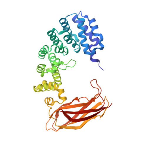
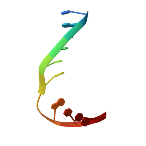
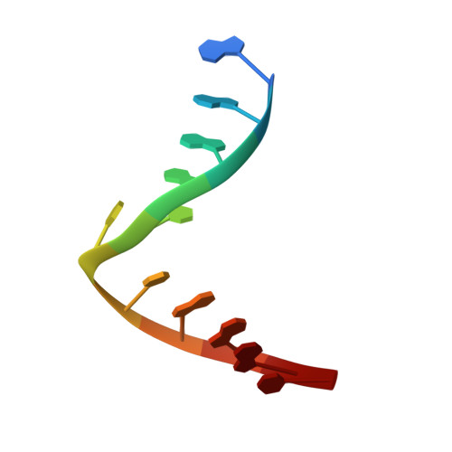
Selective base excision repair of DNA damage by the non-base-flipping DNA glycosylase AlkC.

Shi, R.Mullins, E.A.Shen, X.X.Lay, K.T.Yuen, P.K.David, S.S.Rokas, A.Eichman, B.F.

(2018) EMBO J 37: 63-74

  • DOI: https://doi.org/10.15252/embj.201797833
  • Primary Citation Related Structures: 
    5VHV, 5VI0

  • PubMed Abstract: 

    DNA glycosylases preserve genome integrity and define the specificity of the base excision repair pathway for discreet, detrimental modifications, and thus, the mechanisms by which glycosylases locate DNA damage are of particular interest. Bacterial AlkC and AlkD are specific for cationic alkylated nucleobases and have a distinctive HEAT-like repeat (HLR) fold. AlkD uses a unique non-base-flipping mechanism that enables excision of bulky lesions more commonly associated with nucleotide excision repair. In contrast, AlkC has a much narrower specificity for small lesions, principally N3-methyladenine (3mA). Here, we describe how AlkC selects for and excises 3mA using a non-base-flipping strategy distinct from that of AlkD. A crystal structure resembling a catalytic intermediate complex shows how AlkC uses unique HLR and immunoglobulin-like domains to induce a sharp kink in the DNA, exposing the damaged nucleobase to active site residues that project into the DNA This active site can accommodate and excise N3-methylcytosine (3mC) and N1-methyladenine (1mA), which are also repaired by AlkB-catalyzed oxidative demethylation, providing a potential alternative mechanism for repair of these lesions in bacteria.


  • Organizational Affiliation
    • Department of Biological Sciences, Vanderbilt University, Nashville, TN, USA.

Macromolecule Content 

  • Total Structure Weight: 95.89 kDa 
  • Atom Count: 8,088 
  • Modeled Residue Count: 766 
  • Deposited Residue Count: 766 
  • Unique protein chains: 1
  • Unique nucleic acid chains: 2

Macromolecules


Find similar proteins by:|  3D Structure
Entity ID: 1
MoleculeChains  Sequence LengthOrganismDetailsImage
alkylpurine DNA glycosylase AlkC
A, B
361Pseudomonas fluorescensMutation(s): 0 
Gene Names: PFLU_2162
UniProt
Find proteins for C3K795 (Pseudomonas fluorescens (strain SBW25))
Explore C3K795 
Go to UniProtKB:  C3K795
Entity Groups
Sequence Clusters30% Identity50% Identity70% Identity90% Identity95% Identity100% Identity
UniProt GroupC3K795
Sequence Annotations
Expand
Reference Sequence
Find similar nucleic acids by:  Sequence
Entity ID: 2
MoleculeChains LengthOrganismImage
DNA (5'-D(*TP*GP*TP*CP*CP*AP*(NRI)P*GP*TP*CP*T)-3')
C, E
11synthetic construct
Sequence Annotations
Expand
Reference Sequence
Find similar nucleic acids by:  Sequence
Entity ID: 3
MoleculeChains LengthOrganismImage
DNA (5'-D(*AP*AP*GP*AP*CP*TP*TP*GP*GP*AP*C)-3')
D, F
11synthetic construct
Sequence Annotations
Expand
Reference Sequence

Small Molecules

Ligands 4 Unique
IDChains Name / Formula / InChI Key2D Diagram3D Interactions
9B4

Query on 9B4


Download:Ideal Coordinates CCD File
L [auth D],
O [auth F]
(2R,5R,13R,16R)-9-(hydroxymethyl)-9-{[(2R)-2-hydroxypropoxy]methyl}-5,13-dimethyl-4,7,11,14-tetraoxaheptadecane-2,16-diol
C20 H42 O9
XAZUABIGWFVOAV-FVVUREQNSA-N
MES

Query on MES


Download:Ideal Coordinates CCD File
H [auth A],
I [auth B],
J [auth B]
2-(N-MORPHOLINO)-ETHANESULFONIC ACID
C6 H13 N O4 S
SXGZJKUKBWWHRA-UHFFFAOYSA-N
GOL

Query on GOL


Download:Ideal Coordinates CCD File
G [auth A],
N [auth F]
GLYCEROL
C3 H8 O3
PEDCQBHIVMGVHV-UHFFFAOYSA-N
NA

Query on NA


Download:Ideal Coordinates CCD File
K [auth C],
M [auth E]
SODIUM ION
Na
FKNQFGJONOIPTF-UHFFFAOYSA-N

Experimental Data & Validation

Experimental Data

  • Method: X-RAY DIFFRACTION
  • Resolution: 1.80 Å
  • R-Value Free:  0.164 (Depositor) 
  • R-Value Work:  0.141 (Depositor) 
  • R-Value Observed: 0.142 (Depositor) 
Space Group: P 61
Unit Cell:
Length ( Å )Angle ( ˚ )
a = 198.373α = 90
b = 198.373β = 90
c = 60.189γ = 120
Software Package:
Software NamePurpose
PHENIXrefinement
HKL-2000data reduction
HKL-2000data scaling
PHENIXphasing

Structure Validation

View Full Validation Report


Entry History 

& Funding Information

Deposition Data


Funding OrganizationLocationGrant Number
National Science Foundation (NSF, United States)United States1517695

Revision History  (Full details and data files)

  • Version 1.0: 2017-10-25
    Type: Initial release
  • Version 1.1: 2017-11-01
    Changes: Database references
  • Version 1.2: 2018-01-17
    Changes: Database references
  • Version 1.3: 2019-11-27
    Changes: Author supporting evidence
  • Version 1.4: 2023-10-04
    Changes: Data collection, Database references, Derived calculations, Refinement description