5VFF | pdb_00005vff

Synaptotagmin 1 C2B domain, lead-bound (low occupancy)


Experimental Data Snapshot

  • Method: X-RAY DIFFRACTION
  • Resolution: 1.41 Å
  • R-Value Free: 
    0.191 (Depositor), 0.195 (DCC) 
  • R-Value Work: 
    0.155 (Depositor), 0.157 (DCC) 
  • R-Value Observed: 
    0.158 (Depositor) 

Starting Model: experimental
View more details

wwPDB Validation 3D Report Full Report

Validation slider image for 5VFF

This is version 1.2 of the entry. See complete history

Literature

High affinity interactions of Pb2+with synaptotagmin I.

Katti, S.Her, B.Srivastava, A.K.Taylor, A.B.Lockless, S.W.Igumenova, T.I.

(2018) Metallomics 10: 1211-1222

  • DOI: https://doi.org/10.1039/c8mt00135a
  • Primary Citation Related Structures: 
    5VFE, 5VFF, 5VFG

  • PubMed Abstract: 

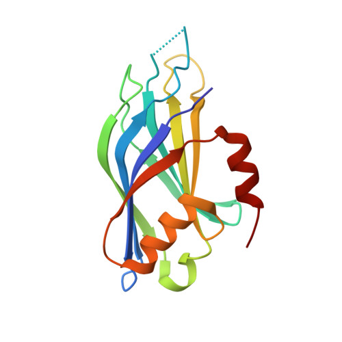
    Lead (Pb) is a potent neurotoxin that disrupts synaptic neurotransmission. We report that Synaptotagmin I (SytI), a key regulator of Ca2+-evoked neurotransmitter release, has two high-affinity Pb2+ binding sites that belong to its cytosolic C2A and C2B domains. The crystal structures of Pb2+-complexed C2 domains revealed that protein-bound Pb2+ ions have holodirected coordination geometries and all-oxygen coordination spheres. The on-rate constants of Pb2+ binding to the C2 domains of SytI are comparable to those of Ca2+ and are diffusion-limited. In contrast, the off-rate constants are at least two orders of magnitude smaller, indicating that Pb2+ can serve as both a thermodynamic and kinetic trap for the C2 domains. We demonstrate, using NMR spectroscopy, that population of these sites by Pb2+ ions inhibits further Ca2+ binding despite the existing coordination vacancies. Our work offers a unique insight into the bioinorganic chemistry of Pb(ii) and suggests a mechanism by which low concentrations of Pb2+ ions can interfere with the Ca2+-dependent function of SytI in the cell.


  • Organizational Affiliation
    • Department of Biochemistry and Biophysics, Texas A&M University, 300 Olsen Boulevard, College Station, TX 77843, USA. tigumenova@tamu.edu.

Macromolecule Content 

  • Total Structure Weight: 17.42 kDa 
  • Atom Count: 1,477 
  • Modeled Residue Count: 149 
  • Deposited Residue Count: 151 
  • Unique protein chains: 1

Macromolecules

Find similar proteins by:|  3D Structure
Entity ID: 1
MoleculeChains  Sequence LengthOrganismDetailsImage
Synaptotagmin-1151Mus musculusMutation(s): 1 
Gene Names: Syt1
UniProt & NIH Common Fund Data Resources
Find proteins for P46096 (Mus musculus)
Explore P46096 
Go to UniProtKB:  P46096
IMPC:  MGI:99667
Entity Groups
Sequence Clusters30% Identity50% Identity70% Identity90% Identity95% Identity100% Identity
UniProt GroupP46096
Sequence Annotations
Expand
Reference Sequence

Small Molecules

Ligands 1 Unique
IDChains Name / Formula / InChI Key2D Diagram3D Interactions
PB

Query on PB


Download:Ideal Coordinates CCD File
B [auth A]LEAD (II) ION
Pb
RVPVRDXYQKGNMQ-UHFFFAOYSA-N

Experimental Data & Validation

Experimental Data

  • Method: X-RAY DIFFRACTION
  • Resolution: 1.41 Å
  • R-Value Free:  0.191 (Depositor), 0.195 (DCC) 
  • R-Value Work:  0.155 (Depositor), 0.157 (DCC) 
  • R-Value Observed: 0.158 (Depositor) 
Space Group: P 21 21 21
Unit Cell:
Length ( Å )Angle ( ˚ )
a = 40.634α = 90
b = 41.117β = 90
c = 82.854γ = 90
Software Package:
Software NamePurpose
PHENIXrefinement
XDSdata reduction
XDSdata scaling
PHASERphasing

Structure Validation

View Full Validation Report


Entry History 

Deposition Data

Revision History  (Full details and data files)

  • Version 1.0: 2018-04-11
    Type: Initial release
  • Version 1.1: 2018-12-19
    Changes: Data collection, Database references
  • Version 1.2: 2023-10-04
    Changes: Data collection, Database references, Derived calculations, Refinement description