5VEJ | pdb_00005vej

High resolution crystal structure of a fluoride-inhibited organo-phosphate-degrading metallohydrolase


Experimental Data Snapshot

  • Method: X-RAY DIFFRACTION
  • Resolution: 1.30 Å
  • R-Value Free: 
    0.144 (Depositor), 0.144 (DCC) 
  • R-Value Work: 
    0.131 (Depositor), 0.131 (DCC) 
  • R-Value Observed: 
    0.131 (Depositor) 

Starting Model: experimental
View more details

wwPDB Validation 3D Report Full Report

Validation slider image for 5VEJ

This is version 1.3 of the entry. See complete history

Literature

High resolution crystal structure of a fluoride-inhibited organophosphate-degrading metallohydrolase.

Selleck, C.Guddat, L.W.Ollis, D.L.Schenk, G.Pedroso, M.M.

(2017) J Inorg Biochem 177: 287-290

  • DOI: https://doi.org/10.1016/j.jinorgbio.2017.06.013
  • Primary Citation Related Structures: 
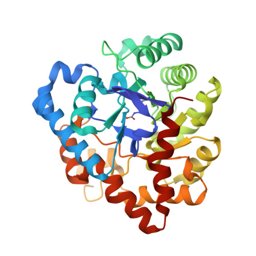
    5VEJ

  • PubMed Abstract: 

    Metal ion-dependent, organophosphate-degrading enzymes (OP hydrolases) have received increasing attention due to their ability to degrade and thus detoxify commonly used pesticides and nerve agents such as sarin and VX. These enzymes thus garner strong potential as bioremediators. The OP hydrolase from Agrobacterium radiobacter (OpdA) is one of the most efficient members of this group of enzymes. Previous studies have indicated that the choice of the hydrolysis-initiating nucleophile may depend on the pH of the reaction, with a metal ion-bridging hydroxide being preferred at lower pH (i.e. pH≤8.5), and a terminally coordinated hydroxide at higher pH (i.e. pH>9.0). Furthermore, fluoride was shown to be a potent inhibitor of the reaction, but only at low pH. Here, the crystal structure (1.3Å, pH6) of OpdA in presence of fluoride is described. While the first coordination sphere in the active site displays minimal changes in the presence of fluoride, the hydrogen bonding network that connects the dimetallic metal center to the substrate binding pocket is disrupted. Thus, the structure of fluoride-inhibited OpdA demonstrates the significance of this hydrogen bond network in controlling the mechanism and function of this enzyme.


  • Organizational Affiliation
    • School of Chemistry and Molecular Biosciences, The University of Queensland, St. Lucia, Queensland 4072, Australia.

Macromolecule Content 

  • Total Structure Weight: 35.84 kDa 
  • Atom Count: 3,098 
  • Modeled Residue Count: 328 
  • Deposited Residue Count: 328 
  • Unique protein chains: 1

Macromolecules

Find similar proteins by:|  3D Structure
Entity ID: 1
MoleculeChains  Sequence LengthOrganismDetailsImage
Phosphotriesterase328Agrobacterium tumefaciensMutation(s): 0 
Gene Names: opdA
UniProt
Find proteins for Q93LD7 (Agrobacterium tumefaciens)
Explore Q93LD7 
Go to UniProtKB:  Q93LD7
Entity Groups
Sequence Clusters30% Identity50% Identity70% Identity90% Identity95% Identity100% Identity
UniProt GroupQ93LD7
Sequence Annotations
Expand
Reference Sequence

Experimental Data & Validation

Experimental Data

  • Method: X-RAY DIFFRACTION
  • Resolution: 1.30 Å
  • R-Value Free:  0.144 (Depositor), 0.144 (DCC) 
  • R-Value Work:  0.131 (Depositor), 0.131 (DCC) 
  • R-Value Observed: 0.131 (Depositor) 
Space Group: P 31 2 1
Unit Cell:
Length ( Å )Angle ( ˚ )
a = 109.072α = 90
b = 109.072β = 90
c = 62.79γ = 120
Software Package:
Software NamePurpose
PHENIXrefinement
PHENIXphasing

Structure Validation

View Full Validation Report


Entry History 

Deposition Data

  • Released Date: 2017-07-19 
  • Deposition Author(s): Selleck, C.

Revision History  (Full details and data files)

  • Version 1.0: 2017-07-19
    Type: Initial release
  • Version 1.1: 2018-01-03
    Changes: Database references
  • Version 1.2: 2023-10-04
    Changes: Data collection, Database references, Derived calculations, Refinement description
  • Version 1.3: 2023-11-15
    Changes: Data collection