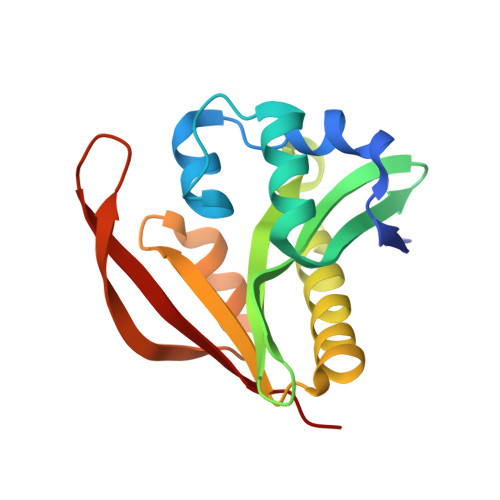
5VD6

Crystal structure of a GNAT superfamily acetyltransferase PA4794 in complex with bisubstrate analog 6


Experimental Data Snapshot

  • Method: X-RAY DIFFRACTION
  • Resolution: 1.20 Å
  • R-Value Free: 0.163 
  • R-Value Work: 0.132 
  • R-Value Observed: 0.134 

wwPDB Validation   3D Report Full Report


Ligand Structure Quality Assessment 


This is version 1.4 of the entry. See complete history


Literature

Generating enzyme and radical-mediated bisubstrates as tools for investigating Gcn5-related N-acetyltransferases.

Reidl, C.Majorek, K.A.Dang, J.Tran, D.Jew, K.Law, M.Payne, Y.Minor, W.Becker, D.P.Kuhn, M.L.

(2017) FEBS Lett 591: 2348-2361

  • DOI: https://doi.org/10.1002/1873-3468.12753
  • Primary Citation of Related Structures:  
    5VD6, 5VDB

  • PubMed Abstract: 

    Gcn5-related N-acetyltransferases (GNATs) are found in all kingdoms of life and catalyze important acyl transfer reactions in diverse cellular processes. While many 3D structures of GNATs have been determined, most do not contain acceptor substrates in their active sites. To expand upon existing crystallographic strategies for improving acceptor-bound GNAT structures, we synthesized peptide substrate analogs and reacted them with CoA in PA4794 protein crystals. We found two separate mechanisms for bisubstrate formation: (a) a novel X-ray induced radical-mediated alkylation of CoA with an alkene peptide and (b) direct alkylation of CoA with a halogenated peptide. Our approach is widely applicable across the GNAT superfamily and can be used to improve the success rate of obtaining liganded structures of other acyltransferases.


  • Organizational Affiliation

    Department of Chemistry, Loyola University Chicago, IL, USA.


Macromolecules
Find similar proteins by:  (by identity cutoff)  |  3D Structure
Entity ID: 1
MoleculeChains Sequence LengthOrganismDetailsImage
acetyltransferase PA4794162Pseudomonas aeruginosa PAO1Mutation(s): 0 
Gene Names: PA4794
UniProt
Find proteins for Q9HV14 (Pseudomonas aeruginosa (strain ATCC 15692 / DSM 22644 / CIP 104116 / JCM 14847 / LMG 12228 / 1C / PRS 101 / PAO1))
Explore Q9HV14 
Go to UniProtKB:  Q9HV14
Entity Groups  
Sequence Clusters30% Identity50% Identity70% Identity90% Identity95% Identity100% Identity
UniProt GroupQ9HV14
Sequence Annotations
Expand
  • Reference Sequence
Small Molecules
Ligands 2 Unique
IDChains Name / Formula / InChI Key2D Diagram3D Interactions
93P
Query on 93P

Download Ideal Coordinates CCD File 
H [auth A](3R,5S,9R,23S)-1-[(2R,3S,4R,5R)-5-(6-amino-9H-purin-9-yl)-4-hydroxy-3-(phosphonooxy)tetrahydrofuran-2-yl]-3,5,9-trihydroxy-8,8-dimethyl-10,14-dioxo-23-({[(phenylacetyl)amino]acetyl}amino)-2,4,6-trioxa-18-thia-11,15-diaza-3,5-diphosphatetracosan-24-oic acid 3,5-dioxide (non-preferred name)
C37 H56 N9 O20 P3 S
OMXLQYGFXWCBIK-IRRLZKHESA-N
SO4
Query on SO4

Download Ideal Coordinates CCD File 
B [auth A]
C [auth A]
D [auth A]
E [auth A]
F [auth A]
B [auth A],
C [auth A],
D [auth A],
E [auth A],
F [auth A],
G [auth A]
SULFATE ION
O4 S
QAOWNCQODCNURD-UHFFFAOYSA-L
Experimental Data & Validation

Experimental Data

  • Method: X-RAY DIFFRACTION
  • Resolution: 1.20 Å
  • R-Value Free: 0.163 
  • R-Value Work: 0.132 
  • R-Value Observed: 0.134 
  • Space Group: P 21 21 2
Unit Cell:
Length ( Å )Angle ( ˚ )
a = 57.088α = 90
b = 76.151β = 90
c = 38.98γ = 90
Software Package:
Software NamePurpose
REFMACrefinement
HKL-3000data scaling
HKL-2000data collection
PDB_EXTRACTdata extraction
MOLREPphasing
HKL-3000data reduction
DENZOdata reduction
SCALEPACKdata scaling

Structure Validation

View Full Validation Report


Ligand Structure Quality Assessment 


Entry History & Funding Information

Deposition Data


Funding OrganizationLocationGrant Number
National Institutes of Health/National Institute of General Medical Sciences (NIH/NIGMS)United StatesU54 GM094585

Revision History  (Full details and data files)

  • Version 1.0: 2017-07-26
    Type: Initial release
  • Version 1.1: 2017-08-23
    Changes: Database references
  • Version 1.2: 2017-11-22
    Changes: Refinement description
  • Version 1.3: 2020-01-01
    Changes: Author supporting evidence
  • Version 1.4: 2023-10-04
    Changes: Data collection, Database references, Refinement description