5V7Z

SSNMR Structure of the Human RIP1/RIP3 Necrosome


Experimental Data Snapshot

  • Method: SOLID-STATE NMR
  • Conformers Calculated: 100 
  • Conformers Submitted: 10 
  • Selection Criteria: structures with the lowest energy 

wwPDB Validation   3D Report Full Report


This is version 1.1 of the entry. See complete history


Literature

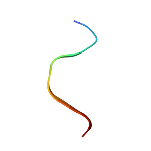
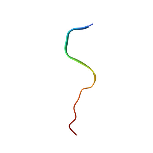
The Structure of the Necrosome RIPK1-RIPK3 Core, a Human Hetero-Amyloid Signaling Complex.

Mompean, M.Li, W.Li, J.Laage, S.Siemer, A.B.Bozkurt, G.Wu, H.McDermott, A.E.

(2018) Cell 173: 1244-1253.e10

  • DOI: https://doi.org/10.1016/j.cell.2018.03.032
  • Primary Citation of Related Structures:  
    5V7Z, 5ZCK

  • PubMed Abstract: 
  • The RIPK1-RIPK3 necrosome is an amyloid signaling complex that initiates TNF-induced necroptosis, serving in human immune defense, cancer, and neurodegenerative diseases. RIPK1 and RIPK3 associate through their RIP homotypic interaction motifs with consensus sequences IQIG (RIPK1) and VQVG (RIPK3) ...

    The RIPK1-RIPK3 necrosome is an amyloid signaling complex that initiates TNF-induced necroptosis, serving in human immune defense, cancer, and neurodegenerative diseases. RIPK1 and RIPK3 associate through their RIP homotypic interaction motifs with consensus sequences IQIG (RIPK1) and VQVG (RIPK3). Using solid-state nuclear magnetic resonance, we determined the high-resolution structure of the RIPK1-RIPK3 core. RIPK1 and RIPK3 alternately stack (RIPK1, RIPK3, RIPK1, RIPK3, etc.) to form heterotypic β sheets. Two such β sheets bind together along a compact hydrophobic interface featuring an unusual ladder of alternating Ser (from RIPK1) and Cys (from RIPK3). The crystal structure of a four-residue RIPK3 consensus sequence is consistent with the architecture determined by NMR. The RIPK1-RIPK3 core is the first detailed structure of a hetero-amyloid and provides a potential explanation for the specificity of hetero- over homo-amyloid formation and a structural basis for understanding the mechanisms of signal transduction.


    Organizational Affiliation

    Department of Chemistry, Columbia University, New York, NY 10027, USA. Electronic address: aem5@columbia.edu.


Macromolecules

Find similar proteins by:  Sequence   |   3D Structure  

Entity ID: 1
MoleculeChainsSequence LengthOrganismDetailsImage
PRO-LEU-VAL-ASN-ILE-TYR-ASN-CYS-SER-GLY-VAL-GLN-VAL-GLY-ASPA, C, E, G15Homo sapiensMutation(s): 0 
Gene Names: RIPK3RIP3
EC: 2.7.11.1
UniProt & NIH Common Fund Data Resources
Find proteins for Q9Y572 (Homo sapiens)
Explore Q9Y572 
Go to UniProtKB:  Q9Y572
PHAROS:  Q9Y572
Entity Groups  
Sequence Clusters30% Identity50% Identity70% Identity90% Identity95% Identity100% Identity
UniProt GroupQ9Y572
Protein Feature View
Expand
  • Reference Sequence

Find similar proteins by:  Sequence   |   3D Structure  

Entity ID: 2
MoleculeChainsSequence LengthOrganismDetailsImage
THR-ILE-TYR-ASN-SER-THR-GLY-ILE-GLN-ILE-GLY-ALA-TYR-ASN-TYR-MET-GLU-ILEB, D, F, H18Homo sapiensMutation(s): 0 
Gene Names: RIPK1RIPRIP1
EC: 2.7.11.1
UniProt & NIH Common Fund Data Resources
Find proteins for Q13546 (Homo sapiens)
Explore Q13546 
Go to UniProtKB:  Q13546
PHAROS:  Q13546
Entity Groups  
Sequence Clusters30% Identity50% Identity70% Identity90% Identity95% Identity100% Identity
UniProt GroupQ13546
Protein Feature View
Expand
  • Reference Sequence
Experimental Data & Validation

Experimental Data

  • Method: SOLID-STATE NMR
  • Conformers Calculated: 100 
  • Conformers Submitted: 10 
  • Selection Criteria: structures with the lowest energy 

Structure Validation

View Full Validation Report


Entry History 

Deposition Data

Revision History  (Full details and data files)

  • Version 1.0: 2018-03-28
    Type: Initial release
  • Version 1.1: 2019-04-10
    Changes: Data collection, Database references