5V61

Phospho-ERK2 bound to bivalent inhibitor SBP2


Experimental Data Snapshot

  • Method: X-RAY DIFFRACTION
  • Resolution: 2.20 Å
  • R-Value Free: 0.212 
  • R-Value Work: 0.176 
  • R-Value Observed: 0.178 

wwPDB Validation   3D Report Full Report


Ligand Structure Quality Assessment 


This is version 1.3 of the entry. See complete history


Literature

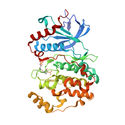
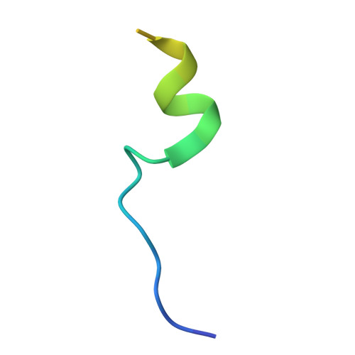
Structure-Guided Strategy for the Development of Potent Bivalent ERK Inhibitors.

Lechtenberg, B.C.Mace, P.D.Sessions, E.H.Williamson, R.Stalder, R.Wallez, Y.Roth, G.P.Riedl, S.J.Pasquale, E.B.

(2017) ACS Med Chem Lett 8: 726-731

  • DOI: https://doi.org/10.1021/acsmedchemlett.7b00127
  • Primary Citation of Related Structures:  
    5V60, 5V61, 5V62

  • PubMed Abstract: 
  • ERK is the effector kinase of the RAS-RAF-MEK-ERK signaling cascade, which promotes cell transformation and malignancy in many cancers and is thus a major drug target in oncology. Kinase inhibitors targeting RAF or MEK are already used for the treatment of certain cancers, such as melanoma ...

    ERK is the effector kinase of the RAS-RAF-MEK-ERK signaling cascade, which promotes cell transformation and malignancy in many cancers and is thus a major drug target in oncology. Kinase inhibitors targeting RAF or MEK are already used for the treatment of certain cancers, such as melanoma. Although the initial response to these drugs can be dramatic, development of drug resistance is a major challenge, even with combination therapies targeting both RAF and MEK. Importantly, most resistance mechanisms still rely on activation of the downstream effector kinase ERK, making it a promising target for drug development efforts. Here, we report the design and structural/functional characterization of a set of bivalent ERK inhibitors that combine a small molecule inhibitor that binds to the ATP-binding pocket with a peptide that selectively binds to an ERK protein interaction surface, the D-site recruitment site (DRS). Our studies show that the lead bivalent inhibitor, SBP3, has markedly improved potency compared to the small molecule inhibitor alone. Unexpectedly, we found that SBP3 also binds to several ERK-related kinases that contain a DRS, highlighting the importance of experimentally verifying the predicted specificity of bivalent inhibitors. However, SBP3 does not target any other kinases belonging to the same CMGC branch of the kinome. Additionally, our modular click chemistry inhibitor design facilitates the generation of different combinations of small molecule inhibitors with ERK-targeting peptides.


    Organizational Affiliation

    Pathology Department, University of California San Diego, La Jolla, California 92093, United States.


Macromolecules
Find similar proteins by:  (by identity cutoff)  |  3D Structure
Entity ID: 1
MoleculeChainsSequence LengthOrganismDetailsImage
Mitogen-activated protein kinase 1A356Homo sapiensMutation(s): 0 
Gene Names: MAPK1ERK2PRKM1PRKM2
EC: 2.7.11.24
UniProt & NIH Common Fund Data Resources
Find proteins for P28482 (Homo sapiens)
Explore P28482 
Go to UniProtKB:  P28482
PHAROS:  P28482
Entity Groups  
Sequence Clusters30% Identity50% Identity70% Identity90% Identity95% Identity100% Identity
UniProt GroupP28482
Protein Feature View
Expand
  • Reference Sequence
Find similar proteins by:  (by identity cutoff)  |  3D Structure
Entity ID: 2
MoleculeChainsSequence LengthOrganismDetailsImage
Ribosomal protein S6 kinase alpha-1,Protein TatB [auth I]28Homo sapiensHIV-1 M:B_HXB2R
This entity is chimeric
Mutation(s): 0 
Gene Names: RPS6KA1tat
EC: 2.7.11.1
UniProt & NIH Common Fund Data Resources
Find proteins for Q15418 (Homo sapiens)
Explore Q15418 
Go to UniProtKB:  Q15418
PHAROS:  Q15418
Find proteins for P04608 (Human immunodeficiency virus type 1 group M subtype B (isolate HXB2))
Explore P04608 
Go to UniProtKB:  P04608
Entity Groups  
Sequence Clusters30% Identity50% Identity70% Identity90% Identity95% Identity100% Identity
UniProt GroupsQ15418P04608
Protein Feature View
Expand
  • Reference Sequence
Small Molecules
Ligands 3 Unique
IDChainsName / Formula / InChI Key2D Diagram3D Interactions
FRZ
Query on FRZ

Download Ideal Coordinates CCD File 
F [auth I]5-(2-PHENYLPYRAZOLO[1,5-A]PYRIDIN-3-YL)-1H-PYRAZOLO[3,4-C]PYRIDAZIN-3-AMINE
C18 H13 N7
XVECMUKVOMUNLE-UHFFFAOYSA-N
 Ligand Interaction
90A
Query on 90A

Download Ideal Coordinates CCD File 
G [auth I]2-oxo-6,9,12,15-tetraoxa-3-azaoctadecan-18-oic acid
C13 H25 N O7
QYCYVPYJQWWUNK-UHFFFAOYSA-N
 Ligand Interaction
GOL
Query on GOL

Download Ideal Coordinates CCD File 
C [auth A],
D [auth A],
E [auth A]
GLYCEROL
C3 H8 O3
PEDCQBHIVMGVHV-UHFFFAOYSA-N
 Ligand Interaction
Modified Residues  2 Unique
IDChainsTypeFormula2D DiagramParent
PTR
Query on PTR
A L-PEPTIDE LINKINGC9 H12 N O6 PTYR
TPO
Query on TPO
A L-PEPTIDE LINKINGC4 H10 N O6 PTHR
Binding Affinity Annotations 
IDSourceBinding Affinity
FRZ BindingDB:  5V61 Ki: min: 310, max: 310 (nM) from 2 assay(s)
Kd: 850 (nM) from 1 assay(s)
IC50: min: 1200, max: 1900 (nM) from 2 assay(s)
Experimental Data & Validation

Experimental Data

  • Method: X-RAY DIFFRACTION
  • Resolution: 2.20 Å
  • R-Value Free: 0.212 
  • R-Value Work: 0.176 
  • R-Value Observed: 0.178 
  • Space Group: P 21 21 21
Unit Cell:
Length ( Å )Angle ( ˚ )
a = 42.217α = 90
b = 78.328β = 90
c = 152.187γ = 90
Software Package:
Software NamePurpose
REFMACrefinement
XDSdata reduction
Aimlessdata scaling
PHASERphasing
CrystalCleardata collection
Cootmodel building

Structure Validation

View Full Validation Report


Ligand Structure Quality Assessment 


Entry History & Funding Information

Deposition Data


Funding OrganizationLocationGrant Number
National Institutes of Health/National Cancer Institute (NIH/NCI)United StatesR01CA160457

Revision History  (Full details and data files)

  • Version 1.0: 2017-07-26
    Type: Initial release
  • Version 1.1: 2017-09-13
    Changes: Author supporting evidence
  • Version 1.2: 2018-04-18
    Changes: Data collection, Database references
  • Version 1.3: 2019-12-04
    Changes: Author supporting evidence