5V5M | pdb_00005v5m

Crystal structure of HLA-B*5701 complex with HIV-1 gag derived peptide TW10


Experimental Data Snapshot

  • Method: X-RAY DIFFRACTION
  • Resolution: 2.88 Å
  • R-Value Free: 
    0.244 (Depositor), 0.232 (DCC) 
  • R-Value Work: 
    0.198 (Depositor), 0.198 (DCC) 
  • R-Value Observed: 
    0.201 (Depositor) 

Starting Model: experimental
View more details

wwPDB Validation 3D Report Full Report

Validation slider image for 5V5M

This is version 1.3 of the entry. See complete history

Literature

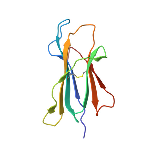
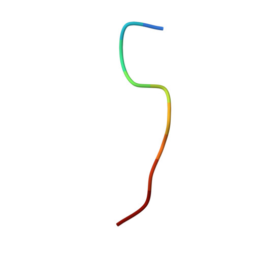
Crystal structure of HLA-B*5801 with a TW10 HIV Gag epitope reveals a novel mode of peptide presentation.

Li, X.Lamothe, P.A.Walker, B.D.Wang, J.H.

(2017) Cell Mol Immunol 14: 631-634

  • DOI: https://doi.org/10.1038/cmi.2017.24
  • Primary Citation Related Structures: 
    5V5L, 5V5M

  • Organizational Affiliation
    • Department of Medical Oncology and Cancer Biology, Dana-Farber Cancer Institute, Harvard Medical School, Boston, MA 02215, USA.

Macromolecule Content 

  • Total Structure Weight: 89.85 kDa 
  • Atom Count: 6,369 
  • Modeled Residue Count: 772 
  • Deposited Residue Count: 774 
  • Unique protein chains: 3

Macromolecules

Find similar proteins by:|  3D Structure
Entity ID: 1
MoleculeChains  Sequence LengthOrganismDetailsImage
HLA class I histocompatibility antigen, B-57 alpha chain
A, C
278Homo sapiensMutation(s): 0 
Gene Names: HLA-BHLAB
UniProt & NIH Common Fund Data Resources
Find proteins for P01889 (Homo sapiens)
Explore P01889 
Go to UniProtKB:  P01889
PHAROS:  P01889
GTEx:  ENSG00000234745 
Entity Groups
Sequence Clusters30% Identity50% Identity70% Identity90% Identity95% Identity100% Identity
UniProt GroupP01889
Sequence Annotations
Expand
Reference Sequence
Find similar proteins by:|  3D Structure
Entity ID: 2
MoleculeChains  Sequence LengthOrganismDetailsImage
Beta-2-microglobulin
B, D
99Homo sapiensMutation(s): 0 
Gene Names: B2MCDABP0092HDCMA22P
UniProt & NIH Common Fund Data Resources
Find proteins for P61769 (Homo sapiens)
Explore P61769 
Go to UniProtKB:  P61769
PHAROS:  P61769
GTEx:  ENSG00000166710 
Entity Groups
Sequence Clusters30% Identity50% Identity70% Identity90% Identity95% Identity100% Identity
UniProt GroupP61769
Sequence Annotations
Expand
Reference Sequence
Find similar proteins by:  Sequence   |   3D Structure  
Entity ID: 3
MoleculeChains  Sequence LengthOrganismDetailsImage
TW10
E, F
10Human immunodeficiency virus 1Mutation(s): 0 
UniProt
Find proteins for P03366 (Human immunodeficiency virus type 1 group M subtype B (isolate BH10))
Explore P03366 
Go to UniProtKB:  P03366
Entity Groups
Sequence Clusters30% Identity50% Identity70% Identity90% Identity95% Identity100% Identity
UniProt GroupP03366
Sequence Annotations
Expand
Reference Sequence

Experimental Data & Validation

Experimental Data

  • Method: X-RAY DIFFRACTION
  • Resolution: 2.88 Å
  • R-Value Free:  0.244 (Depositor), 0.232 (DCC) 
  • R-Value Work:  0.198 (Depositor), 0.198 (DCC) 
  • R-Value Observed: 0.201 (Depositor) 
Space Group: P 21 21 21
Unit Cell:
Length ( Å )Angle ( ˚ )
a = 69.674α = 90
b = 82.622β = 90
c = 157.779γ = 90
Software Package:
Software NamePurpose
PHENIXrefinement
HKL-3000data reduction
HKL-3000data scaling
PHENIXphasing

Structure Validation

View Full Validation Report


Entry History 

Deposition Data

Revision History  (Full details and data files)

  • Version 1.0: 2017-06-14
    Type: Initial release
  • Version 1.1: 2017-07-12
    Changes: Database references
  • Version 1.2: 2023-10-04
    Changes: Data collection, Database references, Refinement description
  • Version 1.3: 2024-11-20
    Changes: Structure summary