5V2S

Crystal structure of glycoprotein B from Herpes Simplex Virus type I

  • Classification: VIRAL PROTEIN
  • Organism(s): Human alphaherpesvirus 1
  • Expression System: Spodoptera frugiperda
  • Mutation(s): No 
  • Membrane Protein: Yes  OPMMemProtMD

  • Deposited: 2017-03-06 Released: 2018-05-16 
  • Deposition Author(s): Cooper, R.S., Heldwein, E.E.
  • Funding Organization(s): National Institutes of Health/National Institute Of Allergy and Infectious Diseases (NIH/NIAID), National Institutes of Health/National Institute of General Medical Sciences (NIH/NIGMS), Burroughs Welcome Fund

Experimental Data Snapshot

  • Method: X-RAY DIFFRACTION
  • Resolution: 3.60 Å
  • R-Value Free: 0.273 
  • R-Value Work: 0.244 
  • R-Value Observed: 0.247 

wwPDB Validation   3D Report Full Report


Ligand Structure Quality Assessment 


This is version 2.1 of the entry. See complete history


Literature

Structural basis for membrane anchoring and fusion regulation of the herpes simplex virus fusogen gB.

Cooper, R.S.Georgieva, E.R.Borbat, P.P.Freed, J.H.Heldwein, E.E.

(2018) Nat Struct Mol Biol 25: 416-424

  • DOI: https://doi.org/10.1038/s41594-018-0060-6
  • Primary Citation of Related Structures:  
    5V2S, 6BM8

  • PubMed Abstract: 

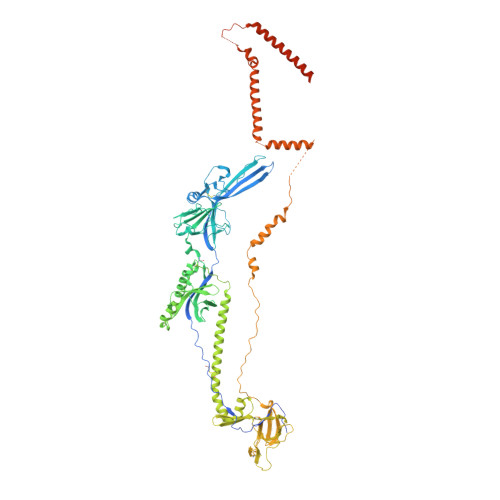
    Viral fusogens merge viral and cell membranes during cell penetration. Their ectodomains drive fusion by undergoing large-scale refolding, but little is known about the functionally important regions located within or near the membrane. Here we report the crystal structure of full-length glycoprotein B (gB), the fusogen from herpes simplex virus, complemented by electron spin resonance measurements. The membrane-proximal (MPR), transmembrane (TMD), and cytoplasmic (CTD) domains form a uniquely folded trimeric pedestal beneath the ectodomain, which balances dynamic flexibility with extensive, stabilizing membrane interactions. The postfusion conformation of the ectodomain suggests that the CTD likewise adopted the postfusion form. However, hyperfusogenic mutations, which destabilize the prefusion state of gB, target key interfaces and structural motifs that reinforce the observed CTD structure. Thus, a similar CTD structure must stabilize gB in its prefusion state. Our data suggest a model for how this dynamic, membrane-dependent 'clamp' controls the fusogenic refolding of gB.


  • Organizational Affiliation

    Department of Molecular Biology and Microbiology, Tufts University School of Medicine, Boston, MA, USA.


Macromolecules
Find similar proteins by:  (by identity cutoff)  |  3D Structure
Entity ID: 1
MoleculeChains Sequence LengthOrganismDetailsImage
Envelope glycoprotein B841Human alphaherpesvirus 1Mutation(s): 0 
Gene Names: UL27HHV1gp041
Membrane Entity: Yes 
UniProt
Find proteins for P10211 (Human herpesvirus 1 (strain 17))
Explore P10211 
Go to UniProtKB:  P10211
Entity Groups  
Sequence Clusters30% Identity50% Identity70% Identity90% Identity95% Identity100% Identity
UniProt GroupP10211
Sequence Annotations
Expand
  • Reference Sequence
Oligosaccharides

Help

Entity ID: 2
MoleculeChains Length2D Diagram Glycosylation3D Interactions
2-acetamido-2-deoxy-beta-D-glucopyranose-(1-4)-2-acetamido-2-deoxy-beta-D-glucopyranose
B, C
2N-Glycosylation
Glycosylation Resources
GlyTouCan:  G42666HT
GlyCosmos:  G42666HT
GlyGen:  G42666HT
Small Molecules
Ligands 1 Unique
IDChains Name / Formula / InChI Key2D Diagram3D Interactions
NAG
Query on NAG

Download Ideal Coordinates CCD File 
D [auth A]2-acetamido-2-deoxy-beta-D-glucopyranose
C8 H15 N O6
OVRNDRQMDRJTHS-FMDGEEDCSA-N
Experimental Data & Validation

Experimental Data

  • Method: X-RAY DIFFRACTION
  • Resolution: 3.60 Å
  • R-Value Free: 0.273 
  • R-Value Work: 0.244 
  • R-Value Observed: 0.247 
  • Space Group: P 3 2 1
Unit Cell:
Length ( Å )Angle ( ˚ )
a = 118.66α = 90
b = 118.66β = 90
c = 216.464γ = 120
Software Package:
Software NamePurpose
PHENIXrefinement
XDSdata reduction
Aimlessdata scaling
PHASERphasing

Structure Validation

View Full Validation Report


Ligand Structure Quality Assessment 


Entry History & Funding Information

Deposition Data


Funding OrganizationLocationGrant Number
National Institutes of Health/National Institute Of Allergy and Infectious Diseases (NIH/NIAID)United States1R21AI107171
National Institutes of Health/National Institute of General Medical Sciences (NIH/NIGMS)United States1F32GM115060
Burroughs Welcome FundUnited StatesInvestigators in the Pathogenesis of Infectious Disease Award

Revision History  (Full details and data files)

  • Version 1.0: 2018-05-16
    Type: Initial release
  • Version 1.1: 2018-10-31
    Changes: Data collection, Database references
  • Version 1.2: 2019-12-11
    Changes: Author supporting evidence
  • Version 2.0: 2020-07-29
    Type: Remediation
    Reason: Carbohydrate remediation
    Changes: Atomic model, Data collection, Derived calculations, Structure summary
  • Version 2.1: 2023-10-04
    Changes: Data collection, Database references, Refinement description, Structure summary