5V07

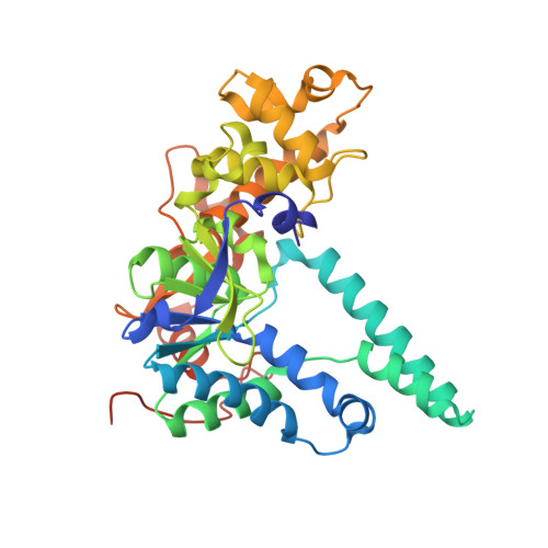
Crystal structure of human exonuclease 1 Exo1 (D173A) in complex with 5' recessed-end DNA (rV)

  • Classification: HYDROLASE/DNA
  • Organism(s): Homo sapiens, synthetic construct
  • Expression System: Escherichia coli
  • Mutation(s): Yes 

  • Deposited: 2017-02-28 Released: 2017-05-24 
  • Deposition Author(s): Shi, Y., Beese, L.S.
  • Funding Organization(s): National Institutes of Health/National Institute of General Medical Sciences (NIH/NIGMS), National Institutes of Health/National Cancer Institute (NIH/NCI)

Experimental Data Snapshot

  • Method: X-RAY DIFFRACTION
  • Resolution: 2.15 Å
  • R-Value Free: 0.220 
  • R-Value Work: 0.176 
  • R-Value Observed: 0.179 

Starting Model: experimental
View more details

wwPDB Validation   3D Report Full Report


This is version 1.5 of the entry. See complete history


Literature

Interplay of catalysis, fidelity, threading, and processivity in the exo- and endonucleolytic reactions of human exonuclease I.

Shi, Y.Hellinga, H.W.Beese, L.S.

(2017) Proc Natl Acad Sci U S A 114: 6010-6015

  • DOI: https://doi.org/10.1073/pnas.1704845114
  • Primary Citation of Related Structures:  
    5UZV, 5V04, 5V05, 5V06, 5V07, 5V08, 5V09, 5V0A, 5V0B, 5V0C, 5V0D, 5V0E

  • PubMed Abstract: 

    Human exonuclease 1 (hExo1) is a member of the RAD2/XPG structure-specific 5'-nuclease superfamily. Its dominant, processive 5'-3' exonuclease and secondary 5'-flap endonuclease activities participate in various DNA repair, recombination, and replication processes. A single active site processes both recessed ends and 5'-flap substrates. By initiating enzyme reactions in crystals, we have trapped hExo1 reaction intermediates that reveal structures of these substrates before and after their exo- and endonucleolytic cleavage, as well as structures of uncleaved, unthreaded, and partially threaded 5' flaps. Their distinctive 5' ends are accommodated by a small, mobile arch in the active site that binds recessed ends at its base and threads 5' flaps through a narrow aperture within its interior. A sequence of successive, interlocking conformational changes guides the two substrate types into a shared reaction mechanism that catalyzes their cleavage by an elaborated variant of the two-metal, in-line hydrolysis mechanism. Coupling of substrate-dependent arch motions to transition-state stabilization suppresses inappropriate or premature cleavage, enhancing processing fidelity. The striking reduction in flap conformational entropy is catalyzed, in part, by arch motions and transient binding interactions between the flap and unprocessed DNA strand. At the end of the observed reaction sequence, hExo1 resets without relinquishing DNA binding, suggesting a structural basis for its processivity.


  • Organizational Affiliation

    Department of Biochemistry, Duke University Medical Center, Durham, NC 27710.


Macromolecules

Find similar proteins by:  (by identity cutoff)  |  3D Structure
Entity ID: 1
MoleculeChains Sequence LengthOrganismDetailsImage
Exonuclease 1A [auth Z]352Homo sapiensMutation(s): 1 
Gene Names: EXO1EXOIHEX1
EC: 3.1
UniProt & NIH Common Fund Data Resources
Find proteins for Q9UQ84 (Homo sapiens)
Explore Q9UQ84 
Go to UniProtKB:  Q9UQ84
PHAROS:  Q9UQ84
GTEx:  ENSG00000174371 
Entity Groups  
Sequence Clusters30% Identity50% Identity70% Identity90% Identity95% Identity100% Identity
UniProt GroupQ9UQ84
Sequence Annotations
Expand
  • Reference Sequence

Find similar nucleic acids by:  Sequence   |   3D Structure  

Entity ID: 2
MoleculeChains LengthOrganismImage
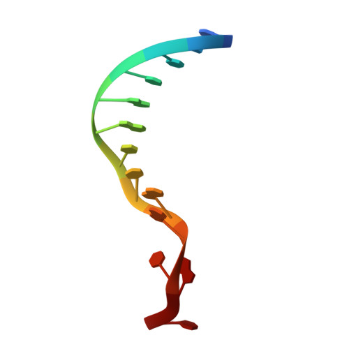
DNA (5'-D(*CP*GP*CP*TP*AP*GP*TP*CP*GP*AP*CP*AP*T)-3')B [auth A]13synthetic construct
Sequence Annotations
Expand
  • Reference Sequence

Find similar nucleic acids by:  Sequence   |   3D Structure  

Entity ID: 3
MoleculeChains LengthOrganismImage
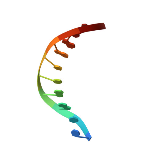
DNA (5'-D(P*TP*CP*GP*AP*CP*TP*AP*GP*CP*G)-3')C [auth B]10synthetic construct
Sequence Annotations
Expand
  • Reference Sequence
Experimental Data & Validation

Experimental Data

  • Method: X-RAY DIFFRACTION
  • Resolution: 2.15 Å
  • R-Value Free: 0.220 
  • R-Value Work: 0.176 
  • R-Value Observed: 0.179 
  • Space Group: P 43 21 2
Unit Cell:
Length ( Å )Angle ( ˚ )
a = 73.837α = 90
b = 73.837β = 90
c = 179.067γ = 90
Software Package:
Software NamePurpose
PHENIXrefinement
HKL-2000data reduction
HKL-2000data scaling
PHASERphasing

Structure Validation

View Full Validation Report


Entry History & Funding Information

Deposition Data


Funding OrganizationLocationGrant Number
National Institutes of Health/National Institute of General Medical Sciences (NIH/NIGMS)United StatesR01 GM091487
National Institutes of Health/National Cancer Institute (NIH/NCI)United StatesP01 CA092584

Revision History  (Full details and data files)

  • Version 1.0: 2017-05-24
    Type: Initial release
  • Version 1.1: 2017-06-07
    Changes: Database references
  • Version 1.2: 2017-06-21
    Changes: Database references
  • Version 1.3: 2017-09-13
    Changes: Author supporting evidence
  • Version 1.4: 2019-12-04
    Changes: Author supporting evidence
  • Version 1.5: 2023-10-04
    Changes: Data collection, Database references, Derived calculations, Refinement description