5UWM

Matrix metalloproteinase-13 complexed with selective inhibitor compound (R)-17a


Experimental Data Snapshot

  • Method: X-RAY DIFFRACTION
  • Resolution: 1.62 Å
  • R-Value Free: 0.178 
  • R-Value Work: 0.129 
  • R-Value Observed: 0.131 

wwPDB Validation   3D Report Full Report


Ligand Structure Quality Assessment 


This is version 1.2 of the entry. See complete history


Literature

Structure-Based Design and Synthesis of Potent and Selective Matrix Metalloproteinase 13 Inhibitors.

Choi, J.Y.Fuerst, R.Knapinska, A.M.Taylor, A.B.Smith, L.Cao, X.Hart, P.J.Fields, G.B.Roush, W.R.

(2017) J Med Chem 60: 5816-5825

  • DOI: https://doi.org/10.1021/acs.jmedchem.7b00514
  • Primary Citation of Related Structures:  
    5UWK, 5UWL, 5UWM, 5UWN

  • PubMed Abstract: 

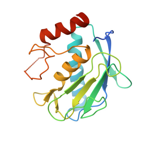
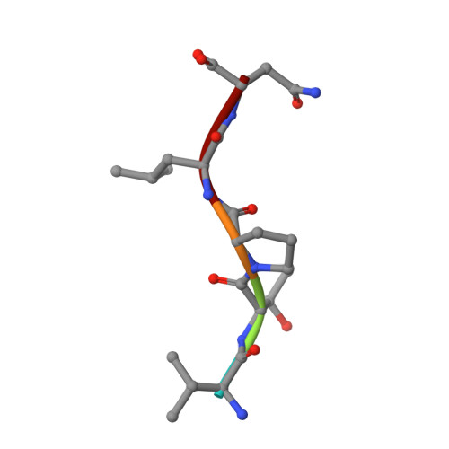
    We describe the use of comparative structural analysis and structure-guided molecular design to develop potent and selective inhibitors (10d and (S)-17b) of matrix metalloproteinase 13 (MMP-13). We applied a three-step process, starting with a comparative analysis of the X-ray crystallographic structure of compound 5 in complex with MMP-13 with published structures of known MMP-13·inhibitor complexes followed by molecular design and synthesis of potent but nonselective zinc-chelating MMP inhibitors (e.g., 10a and 10b). After demonstrating that the pharmacophores of the chelating inhibitors (S)-10a, (R)-10a, and 10b were binding within the MMP-13 active site, the Zn 2+ chelating unit was replaced with nonchelating polar residues that bridged over the Zn 2+ binding site and reached into a solvent accessible area. After two rounds of structural optimization, these design approaches led to small molecule MMP-13 inhibitors 10d and (S)-17b, which bind within the substrate-binding site of MMP-13 and surround the catalytically active Zn 2+ ion without chelating to the metal. These compounds exhibit at least 500-fold selectivity versus other MMPs.


  • Organizational Affiliation

    Department of Chemistry, Scripps Florida , 130 Scripps Way, Jupiter, Florida 33458, United States.


Macromolecules
Find similar proteins by:  (by identity cutoff)  |  3D Structure
Entity ID: 1
MoleculeChains Sequence LengthOrganismDetailsImage
Collagenase 3
A, B
172Homo sapiensMutation(s): 0 
Gene Names: MMP13
EC: 3.4.24
UniProt & NIH Common Fund Data Resources
Find proteins for P45452 (Homo sapiens)
Explore P45452 
Go to UniProtKB:  P45452
PHAROS:  P45452
GTEx:  ENSG00000137745 
Entity Groups  
Sequence Clusters30% Identity50% Identity70% Identity90% Identity95% Identity100% Identity
UniProt GroupP45452
Sequence Annotations
Expand
  • Reference Sequence

Find similar proteins by:  Sequence   |   3D Structure  

Entity ID: 2
MoleculeChains Sequence LengthOrganismDetailsImage
Collagenase 3
C, D
5Homo sapiensMutation(s): 0 
UniProt & NIH Common Fund Data Resources
Find proteins for P45452 (Homo sapiens)
Explore P45452 
Go to UniProtKB:  P45452
PHAROS:  P45452
GTEx:  ENSG00000137745 
Entity Groups  
UniProt GroupP45452
Sequence Annotations
Expand
  • Reference Sequence
Small Molecules
Ligands 3 Unique
IDChains Name / Formula / InChI Key2D Diagram3D Interactions
8OA
Query on 8OA

Download Ideal Coordinates CCD File 
J [auth A],
P [auth B]
(R)-N-(3-methyl-1-(methylamino)-1-oxobutan-2-yl)-5-(4-(((4-oxo-4,5,6,7-tetrahydro-3H-cyclopenta[d]pyrimidin-2-yl)thio)methyl)phenyl)furan-2-carboxamide
C25 H28 N4 O4 S
AHTVZGHNJWEFAC-OAQYLSRUSA-N
ZN
Query on ZN

Download Ideal Coordinates CCD File 
E [auth A],
F [auth A],
K [auth B],
L [auth B]
ZINC ION
Zn
PTFCDOFLOPIGGS-UHFFFAOYSA-N
CA
Query on CA

Download Ideal Coordinates CCD File 
G [auth A]
H [auth A]
I [auth A]
M [auth B]
N [auth B]
G [auth A],
H [auth A],
I [auth A],
M [auth B],
N [auth B],
O [auth B]
CALCIUM ION
Ca
BHPQYMZQTOCNFJ-UHFFFAOYSA-N
Binding Affinity Annotations 
IDSourceBinding Affinity
8OA BindingDB:  5UWM IC50: 356 (nM) from 1 assay(s)
Binding MOAD:  5UWM IC50: 356 (nM) from 1 assay(s)
Experimental Data & Validation

Experimental Data

  • Method: X-RAY DIFFRACTION
  • Resolution: 1.62 Å
  • R-Value Free: 0.178 
  • R-Value Work: 0.129 
  • R-Value Observed: 0.131 
  • Space Group: H 3
Unit Cell:
Length ( Å )Angle ( ˚ )
a = 140.073α = 90
b = 140.073β = 90
c = 46.86γ = 120
Software Package:
Software NamePurpose
PHENIXrefinement
XDSdata reduction
XDSdata scaling
PHASERphasing

Structure Validation

View Full Validation Report


Ligand Structure Quality Assessment 


Entry History 

Deposition Data

Revision History  (Full details and data files)

  • Version 1.0: 2017-07-12
    Type: Initial release
  • Version 1.1: 2017-07-26
    Changes: Database references
  • Version 1.2: 2023-10-04
    Changes: Data collection, Database references, Derived calculations, Refinement description