5UNB | pdb_00005unb

Crystal structure of putative Putative deoxyribonuclease-2 from Burkholderia thailandensis in complex with copper


Experimental Data Snapshot

  • Method: X-RAY DIFFRACTION
  • Resolution: 1.75 Å
  • R-Value Free: 
    0.180 (Depositor), 0.182 (DCC) 
  • R-Value Work: 
    0.148 (Depositor), 0.149 (DCC) 
  • R-Value Observed: 
    0.149 (Depositor) 

Starting Model: experimental
View more details

wwPDB Validation 3D Report Full Report

Validation slider image for 5UNB

This is version 1.3 of the entry. See complete history

Literature

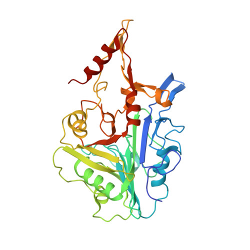
Structure of acid deoxyribonuclease.

Varela-Ramirez, A.Abendroth, J.Mejia, A.A.Phan, I.Q.Lorimer, D.D.Edwards, T.E.Aguilera, R.J.

(2017) Nucleic Acids Res 45: 6217-6227

  • DOI: https://doi.org/10.1093/nar/gkx222
  • Primary Citation Related Structures: 
    5I3E, 5UNB

  • PubMed Abstract: 

    Deoxyribonuclease II (DNase II) is also known as acid deoxyribonuclease because it has optimal activity at the low pH environment of lysosomes where it is typically found in higher eukaryotes. Interestingly, DNase II has also been identified in a few genera of bacteria and is believed to have arisen via horizontal transfer. Here, we demonstrate that recombinant Burkholderia thailandensis DNase II is highly active at low pH in the absence of divalent metal ions, similar to eukaryotic DNase II. The crystal structure of B. thailandensis DNase II shows a dimeric quaternary structure which appears capable of binding double-stranded DNA. Each monomer of B. thailandensis DNase II exhibits a similar overall fold as phospholipase D (PLD), phosphatidylserine synthase (PSS) and tyrosyl-DNA phosphodiesterase (TDP), and conserved catalytic residues imply a similar mechanism. The structural and biochemical data presented here provide insights into the atomic structure and catalytic mechanism of DNase II.


  • Organizational Affiliation
    • Department of Biological Sciences, Border Biomedical Research Center, The University of Texas at El Paso, El Paso, TX 79968, USA.

Macromolecule Content 

  • Total Structure Weight: 79.86 kDa 
  • Atom Count: 6,082 
  • Modeled Residue Count: 681 
  • Deposited Residue Count: 716 
  • Unique protein chains: 1

Macromolecules

Find similar proteins by:|  3D Structure
Entity ID: 1
MoleculeChains  Sequence LengthOrganismDetailsImage
Putative deoxyribonuclease-2
A, B
358Burkholderia thailandensis E264Mutation(s): 0 
Gene Names: BTH_II0389
EC: 3.1
UniProt
Find proteins for Q2T8B0 (Burkholderia thailandensis (strain ATCC 700388 / DSM 13276 / CCUG 48851 / CIP 106301 / E264))
Explore Q2T8B0 
Go to UniProtKB:  Q2T8B0
Entity Groups
Sequence Clusters30% Identity50% Identity70% Identity90% Identity95% Identity100% Identity
UniProt GroupQ2T8B0
Sequence Annotations
Expand
Reference Sequence

Experimental Data & Validation

Experimental Data

  • Method: X-RAY DIFFRACTION
  • Resolution: 1.75 Å
  • R-Value Free:  0.180 (Depositor), 0.182 (DCC) 
  • R-Value Work:  0.148 (Depositor), 0.149 (DCC) 
  • R-Value Observed: 0.149 (Depositor) 
Space Group: P 1 21 1
Unit Cell:
Length ( Å )Angle ( ˚ )
a = 51.55α = 90
b = 61.07β = 90.29
c = 104.47γ = 90
Software Package:
Software NamePurpose
PHENIXrefinement
XDSdata reduction
XSCALEdata scaling
PHASERphasing
Cootmodel building
PDB_EXTRACTdata extraction

Structure Validation

View Full Validation Report


Entry History 

Deposition Data

Revision History  (Full details and data files)

  • Version 1.0: 2017-02-15
    Type: Initial release
  • Version 1.1: 2017-04-12
    Changes: Database references
  • Version 1.2: 2017-06-14
    Changes: Database references
  • Version 1.3: 2023-10-04
    Changes: Data collection, Database references, Derived calculations, Refinement description