5UM6

Crystal Structure of S. pombe Uba1 in a closed conformation


Experimental Data Snapshot

  • Method: X-RAY DIFFRACTION
  • Resolution: 2.79 Å
  • R-Value Free: 0.239 
  • R-Value Work: 0.206 
  • R-Value Observed: 0.207 

wwPDB Validation   3D Report Full Report


Ligand Structure Quality Assessment 


This is version 1.3 of the entry. See complete history


Literature

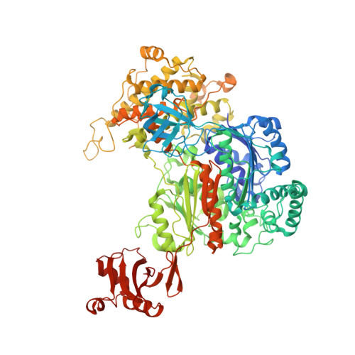
Domain alternation and active site remodeling are conserved structural features of ubiquitin E1.

Lv, Z.Yuan, L.Atkison, J.H.Aldana-Masangkay, G.Chen, Y.Olsen, S.K.

(2017) J Biol Chem 292: 12089-12099

  • DOI: https://doi.org/10.1074/jbc.M117.787622
  • Primary Citation of Related Structures:  
    5UM6

  • PubMed Abstract: 

    E1 enzymes for ubiquitin (Ub) and Ub-like modifiers (Ubls) harbor two catalytic activities that are required for Ub/Ubl activation: adenylation and thioester bond formation. Structural studies of the E1 for the Ubl s mall u biquitin-like mo difier (SUMO) revealed a single active site that is transformed by a conformational switch that toggles its competency for catalysis of these two distinct chemical reactions. Although the mechanisms of adenylation and thioester bond formation revealed by SUMO E1 structures are thought to be conserved in Ub E1, there is currently a lack of structural data supporting this hypothesis. Here, we present a structure of Schizosaccharomyces pombe Uba1 in which the second catalytic cysteine half-domain (SCCH domain) harboring the catalytic cysteine has undergone a 106° rotation that results in a completely different network of intramolecular interactions between the SCCH and adenylation domains and translocation of the catalytic cysteine 12 Å closer to the Ub C terminus compared with previous Uba1 structures. SCCH domain alternation is accompanied by conformational changes within the Uba1 adenylation domains that effectively disassemble the adenylation active site. Importantly, the structural and biochemical data suggest that domain alternation and remodeling of the adenylation active site are interconnected and are intrinsic structural features of Uba1 and that the overall structural basis for adenylation and thioester bond formation exhibited by SUMO E1 is indeed conserved in Ub E1. Finally, the mechanistic insights provided by the novel conformational snapshot of Uba1 presented in this study may guide efforts to develop small molecule inhibitors of this critically important enzyme that is an active target for anticancer therapeutics.


  • Organizational Affiliation

    Department of Biochemistry and Molecular Biology and Hollings Cancer Center, Medical University of South Carolina, Charleston, South Carolina 29425.


Macromolecules
Find similar proteins by:  (by identity cutoff)  |  3D Structure
Entity ID: 1
MoleculeChains Sequence LengthOrganismDetailsImage
Ubiquitin-activating enzyme E1 11,000Schizosaccharomyces pombe 972h-Mutation(s): 1 
Gene Names: ptr3SPBC1604.21cSPBC211.09
EC: 6.2.1.45
UniProt
Find proteins for O94609 (Schizosaccharomyces pombe (strain 972 / ATCC 24843))
Explore O94609 
Go to UniProtKB:  O94609
Entity Groups  
Sequence Clusters30% Identity50% Identity70% Identity90% Identity95% Identity100% Identity
UniProt GroupO94609
Sequence Annotations
Expand
  • Reference Sequence
Small Molecules
Ligands 3 Unique
IDChains Name / Formula / InChI Key2D Diagram3D Interactions
8EA
Query on 8EA

Download Ideal Coordinates CCD File 
C [auth A]N-(2-{[(4-chlorophenyl)methyl]disulfanyl}ethyl)decan-1-amine
C19 H32 Cl N S2
MTCITSZEZJBERN-UHFFFAOYSA-N
8E7
Query on 8E7

Download Ideal Coordinates CCD File 
D [auth A]2-(decylamino)ethane-1-thiol
C12 H27 N S
WOWHRXFAFVDNAP-UHFFFAOYSA-N
SO4
Query on SO4

Download Ideal Coordinates CCD File 
B [auth A]SULFATE ION
O4 S
QAOWNCQODCNURD-UHFFFAOYSA-L
Experimental Data & Validation

Experimental Data

  • Method: X-RAY DIFFRACTION
  • Resolution: 2.79 Å
  • R-Value Free: 0.239 
  • R-Value Work: 0.206 
  • R-Value Observed: 0.207 
  • Space Group: P 21 3
Unit Cell:
Length ( Å )Angle ( ˚ )
a = 145.102α = 90
b = 145.102β = 90
c = 145.102γ = 90
Software Package:
Software NamePurpose
PHENIXrefinement
HKL-2000data reduction
HKL-2000data scaling
PHASERphasing

Structure Validation

View Full Validation Report


Ligand Structure Quality Assessment 


Entry History & Funding Information

Deposition Data


Funding OrganizationLocationGrant Number
National Institutes of Health/National Institute of General Medical Sciences (NIH/NIGMS)United StatesR01 GM115568
National Institutes of Health/National Institute of General Medical Sciences (NIH/NIGMS)United StatesR01 GM086171

Revision History  (Full details and data files)

  • Version 1.0: 2017-06-14
    Type: Initial release
  • Version 1.1: 2017-08-02
    Changes: Author supporting evidence, Database references
  • Version 1.2: 2020-01-01
    Changes: Author supporting evidence
  • Version 1.3: 2023-10-04
    Changes: Data collection, Database references, Refinement description