5ULP

Structure of the NS5 methyltransferase from Zika bound to MS2042


Experimental Data Snapshot

  • Method: X-RAY DIFFRACTION
  • Resolution: 1.55 Å
  • R-Value Free: 0.199 
  • R-Value Work: 0.173 
  • R-Value Observed: 0.174 

wwPDB Validation   3D Report Full Report


Ligand Structure Quality Assessment 


This is version 1.2 of the entry. See complete history


Literature

Development of a S-adenosylmethionine analog that intrudes the RNA-cap binding site of Zika methyltransferase.

Jain, R.Butler, K.V.Coloma, J.Jin, J.Aggarwal, A.K.

(2017) Sci Rep 7: 1632-1632

  • DOI: https://doi.org/10.1038/s41598-017-01756-7
  • Primary Citation of Related Structures:  
    5ULP

  • PubMed Abstract: 

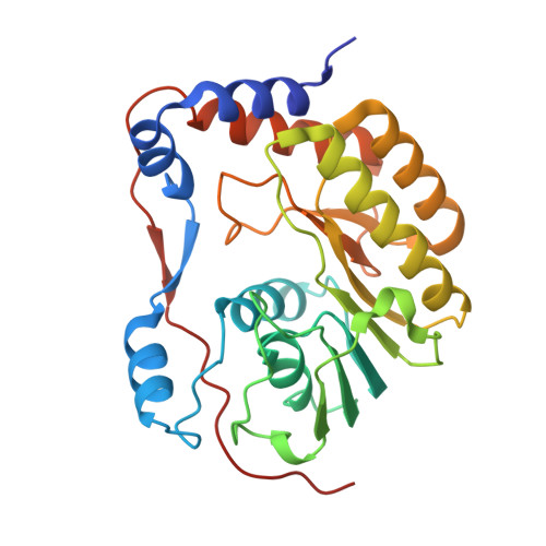
    The Zika virus (ZIKV) has emerged as a major health hazard. We present here a high resolution structure (1.55 Å) of ZIKV NS5 methyltransferase bound to a novel S-adenosylmethionine (SAM) analog in which a 4-fluorophenyl moiety substitutes for the methyl group. We show that the 4-fluorophenyl moiety extends into a portion of the RNA binding tunnel that typically contains the adenosine 2'OH of the RNA-cap moiety. Together, the new SAM analog and the high-resolution crystal structure are a step towards the development of antivirals against ZIKV and other flaviviruses.


  • Organizational Affiliation

    Department of Pharmacological Sciences, Icahn School of Medicine at Mount Sinai, 1425 Madison Avenue, New York, New York, USA.


Macromolecules
Find similar proteins by:  (by identity cutoff)  |  3D Structure
Entity ID: 1
MoleculeChains Sequence LengthOrganismDetailsImage
MRNA cap 0-1 NS5-type methyltransferase
A, B
268Zika virusMutation(s): 0 
UniProt
Find proteins for A0A024B7W1 (Zika virus (isolate ZIKV/Human/French Polynesia/10087PF/2013))
Explore A0A024B7W1 
Go to UniProtKB:  A0A024B7W1
Entity Groups  
Sequence Clusters30% Identity50% Identity70% Identity90% Identity95% Identity100% Identity
UniProt GroupA0A024B7W1
Sequence Annotations
Expand
  • Reference Sequence
Small Molecules
Ligands 5 Unique
IDChains Name / Formula / InChI Key2D Diagram3D Interactions
KB1
Query on KB1

Download Ideal Coordinates CCD File 
C [auth A],
L [auth B]
5'-{[(3S)-3-amino-3-carboxypropyl][(4-fluorophenyl)methyl]amino}-5'-deoxyadenosine
C21 H26 F N7 O5
UBYUMSGWGXUICY-SWQDORGXSA-N
IPA
Query on IPA

Download Ideal Coordinates CCD File 
J [auth A]ISOPROPYL ALCOHOL
C3 H8 O
KFZMGEQAYNKOFK-UHFFFAOYSA-N
URE
Query on URE

Download Ideal Coordinates CCD File 
G [auth A]
H [auth A]
I [auth A]
N [auth B]
O [auth B]
G [auth A],
H [auth A],
I [auth A],
N [auth B],
O [auth B],
P [auth B],
Q [auth B],
R [auth B]
UREA
C H4 N2 O
XSQUKJJJFZCRTK-UHFFFAOYSA-N
CL
Query on CL

Download Ideal Coordinates CCD File 
D [auth A],
E [auth A],
F [auth A],
M [auth B]
CHLORIDE ION
Cl
VEXZGXHMUGYJMC-UHFFFAOYSA-M
NA
Query on NA

Download Ideal Coordinates CCD File 
K [auth A],
S [auth B]
SODIUM ION
Na
FKNQFGJONOIPTF-UHFFFAOYSA-N
Binding Affinity Annotations 
IDSourceBinding Affinity
KB1 Binding MOAD:  5ULP Kd: 2.43e+4 (nM) from 1 assay(s)
Experimental Data & Validation

Experimental Data

  • Method: X-RAY DIFFRACTION
  • Resolution: 1.55 Å
  • R-Value Free: 0.199 
  • R-Value Work: 0.173 
  • R-Value Observed: 0.174 
  • Space Group: P 1 21 1
Unit Cell:
Length ( Å )Angle ( ˚ )
a = 38.989α = 90
b = 111.231β = 93.87
c = 77.4γ = 90
Software Package:
Software NamePurpose
PHENIXrefinement
HKL-2000data reduction
HKL-2000data scaling
Auto-Rickshawphasing

Structure Validation

View Full Validation Report


Ligand Structure Quality Assessment 


Entry History 

Deposition Data

Revision History  (Full details and data files)

  • Version 1.0: 2017-05-17
    Type: Initial release
  • Version 1.1: 2017-05-31
    Changes: Database references
  • Version 1.2: 2023-10-04
    Changes: Data collection, Database references, Derived calculations, Refinement description