5UD5

Crystal structure of the tRNA binding domain of Pyrrolysyl-tRNA synthetase bound to tRNA(Pyl)


Experimental Data Snapshot

  • Method: X-RAY DIFFRACTION
  • Resolution: 2.35 Å
  • R-Value Free: 0.242 
  • R-Value Work: 0.216 
  • R-Value Observed: 0.219 

wwPDB Validation   3D Report Full Report


This is version 1.5 of the entry. See complete history


Literature

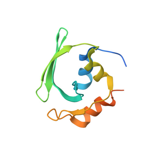
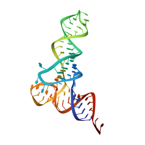
Crystal structures reveal an elusive functional domain of pyrrolysyl-tRNA synthetase.

Suzuki, T.Miller, C.Guo, L.T.Ho, J.M.L.Bryson, D.I.Wang, Y.S.Liu, D.R.Soll, D.

(2017) Nat Chem Biol 13: 1261-1266

  • DOI: https://doi.org/10.1038/nchembio.2497
  • Primary Citation of Related Structures:  
    5UD5, 5V6X

  • PubMed Abstract: 

    Pyrrolysyl-tRNA synthetase (PylRS) is a major tool in genetic code expansion using noncanonical amino acids, yet its structure and function are not completely understood. Here we describe the crystal structure of the previously uncharacterized essential N-terminal domain of this unique enzyme in complex with tRNA Pyl . This structure explains why PylRS remains orthogonal in a broad range of organisms, from bacteria to humans. The structure also illustrates why tRNA Pyl recognition by PylRS is anticodon independent: the anticodon does not contact the enzyme. Then, using standard microbiological culture equipment, we established a new method for laboratory evolution-a noncontinuous counterpart of the previously developed phage-assisted continuous evolution. With this method, we evolved novel PylRS variants with enhanced activity and amino acid specificity. Finally, we employed an evolved PylRS variant to determine its N-terminal domain structure and show how its mutations improve PylRS activity in the genetic encoding of a noncanonical amino acid.


  • Organizational Affiliation

    Department of Molecular Biophysics and Biochemistry, Yale University, New Haven, Connecticut, USA.


Macromolecules

Find similar proteins by:  (by identity cutoff)  |  3D Structure
Entity ID: 1
MoleculeChains Sequence LengthOrganismDetailsImage
Pyrrolysine--tRNA ligase
A, B
109Methanosarcina mazei Go1Mutation(s): 0 
Gene Names: pylSMM_1445
EC: 6.1.1.26
UniProt
Find proteins for Q8PWY1 (Methanosarcina mazei (strain ATCC BAA-159 / DSM 3647 / Goe1 / Go1 / JCM 11833 / OCM 88))
Explore Q8PWY1 
Go to UniProtKB:  Q8PWY1
Entity Groups  
Sequence Clusters30% Identity50% Identity70% Identity90% Identity95% Identity100% Identity
UniProt GroupQ8PWY1
Sequence Annotations
Expand
  • Reference Sequence
Find similar nucleic acids by:  (by identity cutoff)  |  3D Structure
Entity ID: 2
MoleculeChains LengthOrganismImage
RNA (70-MER)
C, D
72Methanosarcina mazei Go1
Sequence Annotations
Expand
  • Reference Sequence
Experimental Data & Validation

Experimental Data

  • Method: X-RAY DIFFRACTION
  • Resolution: 2.35 Å
  • R-Value Free: 0.242 
  • R-Value Work: 0.216 
  • R-Value Observed: 0.219 
  • Space Group: P 31 2 1
Unit Cell:
Length ( Å )Angle ( ˚ )
a = 72.28α = 90
b = 72.28β = 90
c = 238.97γ = 120
Software Package:
Software NamePurpose
PHENIXrefinement
XDSdata reduction
XDSdata scaling
PHENIXphasing

Structure Validation

View Full Validation Report


Entry History & Funding Information

Deposition Data


Funding OrganizationLocationGrant Number
National Institutes of Health/National Institute of General Medical Sciences (NIH/NIGMS)United StatesGM22854

Revision History  (Full details and data files)

  • Version 1.0: 2017-10-11
    Type: Initial release
  • Version 1.1: 2017-10-25
    Changes: Database references
  • Version 1.2: 2017-11-01
    Changes: Database references
  • Version 1.3: 2017-12-06
    Changes: Database references
  • Version 1.4: 2020-01-01
    Changes: Author supporting evidence
  • Version 1.5: 2024-03-06
    Changes: Data collection, Database references