5UBM

Crystal structure of human C1s in complex with inhibitor gigastasin


Experimental Data Snapshot

  • Method: X-RAY DIFFRACTION
  • Resolution: 2.50 Å
  • R-Value Free: 0.222 
  • R-Value Work: 0.177 
  • R-Value Observed: 0.179 

wwPDB Validation   3D Report Full Report


This is version 2.2 of the entry. See complete history


Literature

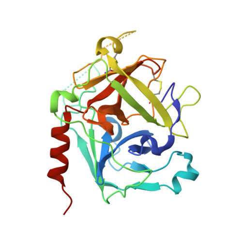
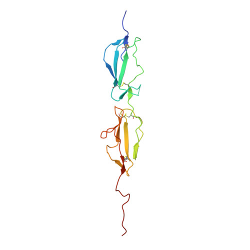
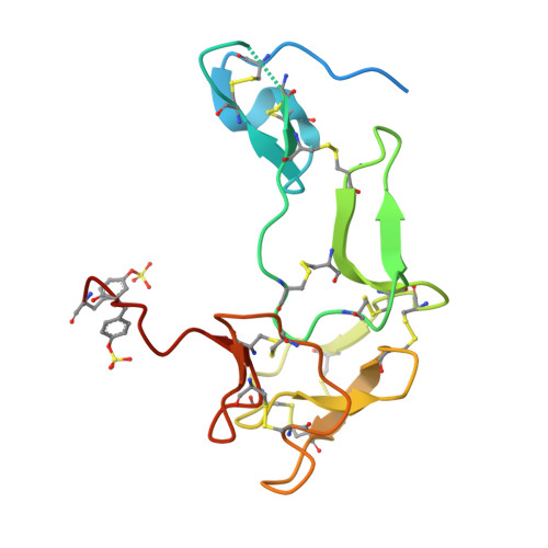
The Structural Basis for Complement Inhibition by Gigastasin, a Protease Inhibitor from the Giant Amazon Leech.

Pang, S.S.Wijeyewickrema, L.C.Hor, L.Tan, S.Lameignere, E.Conway, E.M.Blom, A.M.Mohlin, F.C.Liu, X.Payne, R.J.Whisstock, J.C.Pike, R.N.

(2017) J Immunol 199: 3883-3891

  • DOI: https://doi.org/10.4049/jimmunol.1700158
  • Primary Citation of Related Structures:  
    5UBM

  • PubMed Abstract: 

    Complement is crucial to the immune response, but dysregulation of the system causes inflammatory disease. Complement is activated by three pathways: classical, lectin, and alternative. The classical and lectin pathways are initiated by the C1r/C1s (classical) and MASP-1/MASP-2 (lectin) proteases. Given the role of complement in disease, there is a requirement for inhibitors to control the initiating proteases. In this article, we show that a novel inhibitor, gigastasin, from the giant Amazon leech, potently inhibits C1s and MASP-2, whereas it is also a good inhibitor of MASP-1. Gigastasin is a poor inhibitor of C1r. The inhibitor blocks the active sites of C1s and MASP-2, as well as the anion-binding exosites of the enzymes via sulfotyrosine residues. Complement deposition assays revealed that gigastasin is an effective inhibitor of complement activation in vivo, especially for activation via the lectin pathway. These data suggest that the cumulative effects of inhibiting both MASP-2 and MASP-1 have a greater effect on the lectin pathway than the more potent inhibition of only C1s of the classical pathway.


  • Organizational Affiliation

    Department of Biochemistry and Molecular Biology and Australian Research Council Centre of Excellence in Advanced Molecular Imaging, Biomedicine Discovery Institute, Monash University, Melbourne, Victoria 3800, Australia.


Macromolecules
Find similar proteins by:  (by identity cutoff)  |  3D Structure
Entity ID: 1
MoleculeChains Sequence LengthOrganismDetailsImage
Complement C1s subcomponent252Homo sapiensMutation(s): 0 
EC: 3.4.21.42
UniProt & NIH Common Fund Data Resources
Find proteins for P09871 (Homo sapiens)
Explore P09871 
Go to UniProtKB:  P09871
PHAROS:  P09871
GTEx:  ENSG00000182326 
Entity Groups  
Sequence Clusters30% Identity50% Identity70% Identity90% Identity95% Identity100% Identity
UniProt GroupP09871
Sequence Annotations
Expand
  • Reference Sequence
Find similar proteins by:  (by identity cutoff)  |  3D Structure
Entity ID: 2
MoleculeChains Sequence LengthOrganismDetailsImage
Complement C1s subcomponent152Homo sapiensMutation(s): 0 
EC: 3.4.21.42
UniProt & NIH Common Fund Data Resources
Find proteins for P09871 (Homo sapiens)
Explore P09871 
Go to UniProtKB:  P09871
PHAROS:  P09871
GTEx:  ENSG00000182326 
Entity Groups  
Sequence Clusters30% Identity50% Identity70% Identity90% Identity95% Identity100% Identity
UniProt GroupP09871
Sequence Annotations
Expand
  • Reference Sequence
Find similar proteins by:  (by identity cutoff)  |  3D Structure
Entity ID: 3
MoleculeChains Sequence LengthOrganismDetailsImage
GigastasinC [auth I]137Haementeria ghilianiiMutation(s): 0 
Entity Groups  
Sequence Clusters30% Identity50% Identity70% Identity90% Identity95% Identity100% Identity
Sequence Annotations
Expand
  • Reference Sequence
Oligosaccharides

Help

Entity ID: 4
MoleculeChains Length2D Diagram Glycosylation3D Interactions
2-acetamido-2-deoxy-beta-D-glucopyranose-(1-4)-2-acetamido-2-deoxy-beta-D-glucopyranoseD [auth C]2N-Glycosylation
Glycosylation Resources
GlyTouCan:  G42666HT
GlyCosmos:  G42666HT
GlyGen:  G42666HT
Small Molecules
Modified Residues  1 Unique
IDChains TypeFormula2D DiagramParent
TYS
Query on TYS
C [auth I]L-PEPTIDE LINKINGC9 H11 N O6 STYR
Experimental Data & Validation

Experimental Data

  • Method: X-RAY DIFFRACTION
  • Resolution: 2.50 Å
  • R-Value Free: 0.222 
  • R-Value Work: 0.177 
  • R-Value Observed: 0.179 
  • Space Group: P 31 2 1
Unit Cell:
Length ( Å )Angle ( ˚ )
a = 89.35α = 90
b = 89.35β = 90
c = 146.871γ = 120
Software Package:
Software NamePurpose
PHENIXrefinement
MOSFLMdata reduction
SCALAdata scaling
Blu-Icedata collection

Structure Validation

View Full Validation Report


Entry History 

Deposition Data

Revision History  (Full details and data files)

  • Version 1.0: 2017-11-08
    Type: Initial release
  • Version 1.1: 2017-11-29
    Changes: Database references
  • Version 2.0: 2020-07-29
    Type: Remediation
    Reason: Carbohydrate remediation
    Changes: Atomic model, Data collection, Derived calculations, Structure summary
  • Version 2.1: 2023-10-04
    Changes: Data collection, Database references, Refinement description, Structure summary
  • Version 2.2: 2023-11-15
    Changes: Data collection